ch8-pregnancy

Pregnancy in Sea Otters

Few reports provide comprehensive data concerning the rate of success for delivery and rearing of pups by wild sea otters. It has been estimated that only 30% of pups survive their first year (Jameson and Johnson, 1993; Reidman and Estes, 1990). Immature females are considered less capable than experienced adults in meeting the constant demands imposed by feeding and grooming of newborn pups.

During the wildlife rehabilitation program following the EVOS, nearly 70% of the captured sea otters were female; forty-nine of these were diagnosed as pregnant during admission or at necropsy. In forty (81%) of these females, pregnancy was terminated by: 1) death of the mother (n = 19); 2) abortion of a near term fetus or stillbirth (n = 9); or 3) death of the newborn pup (n = 12). Thirteen newborn pups were transferred to a nursery after their mothers were unable to care for them; all but one of these pups died. Four pregnant females completed the rehabilitation process and were released before delivery. Only five of the pregnant otters were able to deliver and successfully care for live pups in the rehabilitation center (Tuomi et al., 1991).

Mortality in pregnant otters reached 38% and was 4% higher than the overall mortality of captured otters following the EVOS. Pregnant otters admitted during the first three weeks exhibited the greatest degree of oiling and suffered the highest mortality. This was similar to results reported for all sea otters in the rehabilitation program (Williams et al., 1990). Hepatic and renal lipidosis was 2.6 times more common in females than males. Females may have been predisposed to these disorders by the high energy demands of pregnancy and lactation (Lipscomb et al., 1993).

Stillborn pups ranged from 1.2-2.0 kg in body weight; five of these pups showed lesions suggesting death in utero before the onset of labor. Neonatal death (pup survival less than three days) occurred in ten of the liveborn pups; eight of these pups weighed less than 1.4 kg and may have been born prematurely. Samples of tissue from a deceased pup and colostrum from one of the females revealed significant levels of petroleum hydrocarbons (see below). One pup had a large umbilical defect with evisceration and died within a few minutes. Another pup underwent successful surgical repair of an umbilical hernia at two days of age. Uterine torsion was diagnosed postmortem in three females with very large full-term fetuses (fetal weight = 2.0, 2.3, and 2.5 kg).

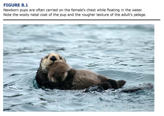
Nine females brought to rehabilitation centers were accompanied by live pups ranging in age from a few days to several months. Two of the nursing females died and their pups were moved to a nursery along with three more pups from this group whose mothers could not care for them. Two of these five separated pups died.

Eleven other orphaned pups were captured and brought into the centers. Orphan pups from the wild had a lower mortality than pups born in the rehabilitation centers. Ten (91%) of the original eleven wild orphaned pups survived and were transferred to seaquaria.

In comparison to the sea otters from the EVOS, reports from a fifteen-year captive breeding program at the Vancouver Aquarium indicate a 72% success rate with eight pups raised to weaning. One stillbirth, one neonatal death, and one pup lost due to lack of maternal care occurred in this series.

ch8-anesthesia

Anesthesia and Physical Restraint

Data concerning the physiological changes occurring with gestation and parturition indicate an increased health risk for pregnant mammals during anesthesia (Table 8.1; Benson and Thurmon, 1987). For example, cardiac work is increased during pregnancy, thus creating a decreased cardiac reserve. The increase in plasma volume may be greater than increases in red blood cell mass, resulting in decreased hemoglobin concentration and packed cell volume (PCV) in the pregnant anima1. If these changes are exacerbated by Heinz body formation associated with petroleum hydrocarbon toxicosis, the pregnant female may become anemic. Struggling during restraint or anesthetic induction could result in pulmonary congestion and heart failure in pregnant animals. The functional residual capacity of the lungs is decreased by pressure on the diaphragm and abdominal organs by the gravid uterus. The pressure of the enlarged uterus on the diaphragm and major abdominal vessels can alter oxygenation and return blood flow especially in marine mammals whose natural buoyancy probably minimizes these effects in the aquatic environment. Hypoxemia and hypercapnia may occur if the animal does not breathe properly. The fetus may be adversely affected by abnormal maternal acid/base balance or by decreases in maternal blood pressure which may reduce blood flow to the placenta and cause fetal hypoxia. Vomiting and aspiration are also more likely to occur in the pregnant animal during anesthesia. Complications include asphyxiation and pneumonitis.

Table8.1

In view of the above complicating factors, there is no ideal anesthetic for pregnant sea otters. Primary consideration should be given to minimizing the time from induction to recovery, and minimizing the effects of depression on the cardiopulmonary and respiratory systems of the mother (Benson and Thurmon, 1987).
The narcotic, fentanyl, was the most commonly used anesthetic at sea otter rehabilitation centers during the EVOS. It was usually combined with diazepam (to reduce the occurrence of seizures) and a tranquilizer such as azaperone or acetylpromazine (see Chapter 3). This protocol had the advantage of rapid and reliable induction after intramuscular injection. In addition, naloxone could be used as a reversal agent after completion of washing or treatments. All of these agents can cross the placenta (Briggs et a1., 1983).

Opiates can cause fetal depression proportional to the degree of analgesia produced. Fetal elimination of opiates may take up to two to six days in some species. Because opiate antagonists such as naloxone also cross the placenta, initial reversal of this depression will occur. However, naloxone itself can cause mild neonatal depression. Because naloxone has a short duration of action, renarcotization from the fentanyl may occur once the naloxone is metabolized and excreted. All otters, and especially neonates delivered within two days of maternal anesthesia, should be monitored for recurring signs of narcosis. Supplemental naloxone should be administered, if indicated.

Phenothiazine tranquilizers, including acetylpromazine, may promote opiateinduced depression but add little to analgesia. They induce hypotension, respiratory depression, and thermal instability. In contrast to the opiates, the duration of action of these tranquilizers is long (lasting up to eight hours) and cannot be reversed. The use of these tranquilizers in pregnant animals should be limited to markedly apprehensive or excited females. The doses should be minimal to produce a calming effect, but not undue generalized depression.

When administered to pregnant animals, diazepam can produce lethargy, hypothermia, and hypotonus in the newborn. These effects are dose related and do not appear to cause severe problems when minimal doses are used (Benson and Thurmon, 1987). As a cautionary note, fetal deformities have been associated with the use of diazepam in the first trimester in human patients (Briggs et a1., 1983).

Inhalation anesthetics such as isoflurane and halothane readily cross the placenta, resulting in rapid fetal and maternal equilibrium. Consequently, a degree of depression is created in the fetus that is proportional to the depth of anesthesia in the mother. Deep levels of maternal anesthesia may cause maternal hypotension, decreased uterine blood flow and fetal acidosis. The same responses are observed in mothers suffering from circulatory collapse due to hypoglycemia, hypothermia, or shock. In view of this, the use of inhalation anesthetic agents in hypotensive pregnant otters should be minimized or avoided.

Sea otters in advanced pregnancy should be placed in lateral recumbency (on their side) during anesthesia to reduce pressure on the diaphragm and major vessels. Anaesthetic times should be kept short. Intravenous or subcutaneous fluids (normal saline or a 1-to-1 mixture of 5% dextrose solution and normal saline; 20 ml/kg/day) should be administered to maintain adequate blood pressure and perfusion. If I available, assisted ventilation or supplemental oxygen may be beneficial in severely depressed animals. However, positive pressure delivery systems are not recommended if interstitial or subcutaneous emphysema is suspected. (See Chapter 5.) Constant monitoring of body temperature is vital and methods to warm or cool the animals must be provided. Care must be exercised when moving anesthetized otters in advanced pregnancy to avoid abrupt rotational movements which might induce uterine torsion.

ch8-medical

Medical Considerations

Abortion has been observed in many species as a response to severe physiological stress. Starvation may result in abortion as a protective mechanism to conserve the maternal animal’s own body reserves. Stresses associated with transport, close housing, sudden reduction or change in food and water intake, and other debilitating factors will cause abortion in pregnant mares, especially during the middle of pregnancy (Roberts, 1980). Miller (1980) notes that abortion in emaciated cattle may occur, but that abortion or premature delivery in these cases does not preserve the life of the anima1. Rather, it heralds a terminal event. The report recommends the induction of parturition at an earlier stage in these animals.

Glucocorticoids administered in repeated doses over several days have been used to stimulate abortion in the last trimester of pregnancy in horses (Roberts, 1980). In contrast, similar treatments have no effect on the termination of pregnancy in many other species. Administration of exogenous adrenocorticotrophin (ACTH) or corticosteroids can disrupt implantation and fetal development in sheep and rats. Currently, it is difficult to predict the concentration of adrenal hormones released during a specific stressful event, and whether the levels would be sufficient to result in abortion (Moberg, 1985).

Several forms of glucocorticoids (dexamethasone, prednisolone, triamcinolone) were routinely used to combat shock in debilitated sea otters during the EVOS. Use of these drugs in females was discontinued after it was suspected that corticosteroids contributed to the high incidence of abortion and stillbirths in the centers. To evaluate this, we compared the outcome of pregnancies for otters receiving and not receiving steroids during rehabilitation (Table 8.2). The total number of otters in this study was relatively small and many other variables influenced the condition of the pregnant animals. In general, survival rates were comparatively better for females receiving corticosteroids at admission. The rates of stillbirths and neonatal deaths were similar for both groups. Based on these results, corticosteroid administration should be considered.

Table8.2

During the EVOS, live pups were taken from twelve females which were unable to care for them during the first few hours after birth. Most of these neonates were never able to nurse and were hypothermic due to the inability of the mother to keep the pup’s fur properly groomed. Newborn pups are susceptible to chilling and may drown or be injured if the female is inattentive. Pups which become hypothermic due to poor grooming or immersion in water are very difficult to stabilize. Only one of the pups removed from its mother survived. It was transferred to Point Defiance Zoo and Aquarium where it died at about four months of age. The poor viability of pups born to debilitated otters at the centers is not surprising, but the effect of oil exposure, capture stress, medical treatments, husbandry techniques, and natural maternal abilities on survival remain uncertain. Although maternal survivorship dramatically improved once the oil weathered, the mortality in the offspring remained high. Viable pups were not delivered in the rehabilitation centers until nine weeks after the oil spill. Fifty percent of the deliveries after this period still resulted in death of the offspring, although no further maternal deaths occurred.

Dystocia (delayed or difficult delivery of a full term fetus) is a potential problem in a rehabilitation center. At least two sea otters treated during the EVOS experienced difficult labor. Oxytocin and calcium injections were administered to one of these females in an effort to strengthen contractions, but the effect was questionable. Both otters eventually delivered very large, stillborn pups following more than twenty-four hours of labor. The untreated female subsequently developed a purulent vaginal discharge and was given antibiotic injections for several days before making a full recovery. A captive sea otter housed for more than two years in a seaquarium had to be assisted with the delivery of a large, stillborn fetus after several hours of unproductive effort. This otter sustained pelvic trauma which resulted in paralysis; death occurred several days later (Vancouver Aquarium Animal Care Department, personal communication). Cesarean section may be performed using fentanyl/diazepam sedation and standard surgical procedures if dystocia is determined to be life threatening.

Uterine torsion has not been reported as a cause of mortality in wild sea otters but has been observed and surgically corrected in an otter housed in a seaquarium (T. D. Williams, Monterey Bay Aquarium; personal communication). The high incidence of uterine torsion observed during the EVOS was a definite concern. The weight of the gravid uterus may not be properly supported when the otter is out of water; transport for prolonged periods in kennel cages while the otter struggles may result in torsion. Excessive rolling and violent activity during capture or while under sedation may also predispose the female to this condition. Female otters in advanced pregnancy should be moved with care and observed closely for signs of abdominal distress. Early surgical intervention is required for correction once torsion has occurred. Prevention or early detection is important.

ch8-husbandry

Husbandry Considerations

Oiled females that have recovered sufficiently to swim and groom normally prior to delivery appear to have the greatest success in raising a pup. Pregnant otters that are unoiled or are captured more than eight weeks following a spill are also more likely to successfully deliver and raise a pup. These otters should be handled as little as possible, housed in large seawater pools, fed, and observed regularly according to standard husbandry protocols (see Chapter 7). The holding pool may house other compatible females, but these animals may have to be removed after delivery if they interfere with the mother in feeding or caring for her pup, or if care of the other otters disturbs the new mother. Pup stealing has been observed frequently in captive sea otters and may create problems if the mother is inexperienced or too debilitated to protect the pup from such interference.

Most otters deliver after relatively short periods of obvious labor, regardless of whether they are in the water or on a dry surface. Some females with free access to water choose to complete their labor on haul out areas. The onset of labor is frequently signaled by a sudden loss of appetite and increased attention to grooming of the vaginal area. Some females appear to rub their lower abdomen which may assist contractions, and most will alternate periods of straining on the haul out with frequent periods of swimming, including vigorous rolling and grooming behaviors. After delivery, the female usually floats on her back in the water and holds the newborn pup on her chest while she licks and dries its fur (Figure 8.1). The placenta will usually be observed trailing from the vaginal opening for up to two hours after delivery. Eventual passage of these membranes appears to cause the female no distress. A moderate bloody discharge also may be observed during the first post partum day, but is seldom noted after that time.

Fig8.1

Once the mother has completed grooming and drying her newborn, a healthy pup will usually move onto the female’s lower abdomen and nurse. The pup will remain nursing and sleeping on its mother’s upturned chest and abdomen for the next three to four weeks, except for short periods when she places the pup on a haulout or leaves it floating on the surface of the water while she feeds or grooms. A pup will vocalize loudly during these short separations and will gradually start to perform simple swimming movements, beginning with the ability to roll onto its back.

Haulouts should be designed to allow females to crawl out of the water without dragging the pup against a sharp edge. Pups can become trapped in overflow outlets or under haulouts unless such structures are adequately enclosed. Pools designed to hold young pups and their mothers should have haul out space which can be gated to separate the animals without undue stress while the pool is cleaned and disinfected. Alternatively, floating maternity pens can be constructed in the prerelease facility. (See Chapter 12 for a description of the different types of enclosures.)

Stillborn pups may be groomed and carried for several hours before the female loses interest. Females may become frantic when handlers attempt to remove weak or dead pups. As a result, a pup may be drowned or crushed if the female will not abandon it. Female otters which are unable to float on the water due to poor coat condition or medical problems can perform pup grooming while in a dry cage. However, they will probably be unable to successfully nurse their offspring.

Once a healthy pup has become bonded to its mother, short separations for purposes of medical treatment or movement within the rehabilitation center cause few problems. When mothers and pups must be separated during transport, it is preferable to place cages so that they may remain in visual contact. In at least one case, a three-month-old pup separated from its mother for medical reasons for more than seven weeks was able to reestablish the maternal bond within twenty-four hours after its return to the mother (J. McBain, Sea World; personal communication).

The high rate of neonatal death can be extremely distressing to the husbandry staff. This problem should be anticipated and addressed during training with special attention to protocols for handling otters whose pups die. Only one or two people should be allowed in the vicinity of any newborn and its mother. Movement of animals for treatment or pen cleaning should be postponed as long as possible, and then performed efficiently by well-trained handlers. In view of the poor survival rate of pups born to otters early in a rehabilitation program, it may be preferable to allow the female otter to care for its pup with minimal interference. A pup that dies in this circumstance can be removed after the mother abandons it without creating undue stress to the female or the staff.

If a decision is made to remove a pup and care for it in a nursery, it may be preferable to do so before the pup nurses; females exposed to crude oil may transfer petroleum hydrocarbons through the colostrum (see following section). In these cases, the benefits of nursing must be balanced against the potential exposure to petroleum hydrocarbons in the mother’s milk. When removing a pup, the mother should be distracted with food or otherwise separated from her pup while a second handler approaches and removes the pup with a net.

Lactating female sea otters have been observed to adopt young pups in a variety of situations (Reidman and Estes, 1990). The successful raising of a pup by a surrogate female in captivity has been reported. This deserves further investigation as an alternative to nursery care for orphaned or abandoned pups.

Lactating females will have a higher nutritional requirement than non-lactating otters (see Chapter 7 for normal dietary requirements). Food should be offered in larger amounts and at more frequent intervals throughout the day (up to 50% body weight per twenty-four hours).

ch8-toxicological

Toxicological Considerations

Toxic substances can pass to a developing fetus through the blood and by simple diffusion across the placental membranes. Currently, it is uncertain whether the placenta can actively prevent the transfer of toxicants from the mother to the fetus. However, the placenta has demonstrated biotransformation properties which could reduce exposure of some substances to the fetus (Klaassen and Rozman, 1991). The effect of an absorbed toxicant will depend on the ability of fetal tissue to concentrate the specific substance. For example, in laboratory mammals the livers of newborns and fetuses will not accumulate some foreign substances and will show much reduced toxicant levels in comparison to maternal liver tissue (Klaassen and Rozman, 1991). Because of a poorly developed blood-brain barrier, the fetal brain may be more susceptible to some toxicants.

In mammals, the effect of a toxicant on the fetus will also depend on the stage of development when exposure occurs (Manson and Wise, 1991). The difference between reversible and irreversible damage often is measured in days. Irreversible effects may be lethal (abortion or stillbirth is induced) or nonlethal (retarded or delayed growth of specific organ systems).

As stated in Chapter 1, it is difficult to verify tissue damage from exposure to crude oil in marine mammals. To date, chlorinated hydrocarbons (DDT, PCBs) have been implicated in reproductive disorders in pinnipeds (St. Aubin, 1990). Little information is available on the direct toxicological effects of petroleum hydrocarbons on the reproductive system, pregnancy, or fetal development. Following the EVOS, petroleum hydrocarbon concentrations were measured in the tissues of a newborn pup from a lightly oiled female sea otter in the rehabilitation center. Polycyclic aromatic hydrocarbons (PAHs) were found in the lungs, liver, and kidneys of the pup. Accumulation was especially marked in the kidney, which showed a mean PAH concentration that was three times the levels in other tissues (T. M. Williams, unpublished data). Although mortality of the pup may have been independent of oil contamination, maternal transfer of petroleum hydrocarbons to the developing fetus appears possible.

In mammals, some toxic agents may be transferred to milk by simple diffusion across blood vessels in the mammary glands. This, in turn, creates a pathway of exposure to the nursing animal. The high lipid content of milk promotes the accumulation of lipid-soluble toxicants including many petroleum hydrocarbon compounds. The problem is exacerbated in marine mammals, which have an exceptionally high concentration of lipids in their milk (Worthy, 1990). Colostrum (the secretion of the mammary glands immediately following parturition) contains an even higher lipid content than milk. Species differences in the excretion of lipid-soluble toxicants through milk will depend on the proportion of milk fat derived from the circulation versus milk synthesis in mammary tissue (Klaassen and Rozman, 1991).

Although the results are not conclusive, milk transfer of petroleum hydrocarbons has been implicated for both pinnipeds and sea otters. St. Aubin (1990) suggests that the effects of petroleum hydrocarbon exposure through nursing is heightened in immature animals due to their comparatively low levels of detoxifying enzymes. A colostrum sample taken from a heavily oiled sea otter showed high levels of total paraffinic hydrocarbons (941 ppm; T. M. Williams, unpublished data). Ideally, it would be beneficial to analyze the milk of oiled marine mammals before allowing newborns to nurse in rehabilitation centers.

ch8-summary

Summary

he stresses associated with oil contamination and subsequent rehabilitation may be exacerbated in pregnant otters. As a result, the rehabilitation team must be aware of the unique requirements associated with sedating, treating, housing, and feeding this group of animals. Maternal, placental, and fetal compartments are in close communication; thus, oil contamination and the rehabilitation process also may affect the developing fetus. Maternal transfer of petroleum hydrocarbons to the pup may continue after parturition in the form of contaminants transferred through milk.

Rehabilitators must decide whether to separate newborn pups shortly after birth, especially if additional toxic exposure is eminent from nursing. Pups can be successfully raised in nurseries, but are usually unsuited for release back to the wild. Each pup-mother pair must be evaluated individually depending on degree, type and time of oil exposure of the female, the availability of facilities and personnel for rehabilitation, and plans for eventual disposition of rehabilitated animals.

ch8-lit

Literature Cited

Benson, G. J., and J. C. Thurmon. 1987. “Special anesthetic considerations for cesarian section.” In Principles and practice of veterinary anesthesia. C. E. Short, ed., 337-47. Baltimore: Williams and Wilkins.

Briggs, G. B., T. W. Boendorier, R. K. Freeman, and S. J. Yaffe. 1983. Drugs in Pregnancy and Lactation. Baltimore: Williams and Wilkins.

Engelhardt, F. R. 1983. Petroleum Effects on Marine Mammals. Aquatic Toxicology 4:199-217.
Jameson, R. J., and A. M. Johnson. 1993. Reproductive characteristics of female sea otters. Marine Mammal Science 9 (2): 156-67.

Klaassen, C. D., and K. Rozman. 1991. “Absorption, distribution, and excretion of toxicants.” In Toxicology: The basic science of poisons. M. O. Amdur, J. Doull and C. D. Klaassen, eds., 50-87. New York: Pergamon Press.

Lipscomb, T. P, R. K. Harris, R. B. Moeller, J. M. Pletcher, R. J. Haebler, and B.E. Ballachey. 1993. Histopathological lesions in sea otters exposed to crude oil. Veterinary Pathology 30:1-11.

Manson, J. M., and L. D. Wise. 1991. “Teratogens.” In Toxicology: The basic science of poisons. M. O. Amdur, J. Doull and C. D. Klaassen, eds., 226-56. New York: Pergamon Press.

Miller, R. B. 1980. “Abortion.” In Current therapy in theriogenology. D. A. Morrow, ed., 213-22. Philadelphia: W. B. Saunders.

Moberg, G. P. 1985. “Influence of stress on reproduction: Measure of well being.” In Animal Stress. G. P. Moberg, ed., 245-51. Bethesda: American Physiological Society.

Reidman, M. L., and J. A. Estes. 1990. “Reproduction.” In Sea otter, Enhydra lutris: ecology, behavior, and natural history. U.S. Fish and Wildlife Service Biological Report 90 (14): 59-72.

Roberts, S. J. 1980. “Abortion and other diseases of gestation in mares.” In Current therapy in theriogenology. D. A Morrow, ed., 748-53. Philadelphia: W. B. Saunders.

St. Aubin, D. J. 1990. “Physiologic and toxic effects on pinnipeds.” In Sea mammals and oil: Confronting the risks. J. R. Geraci and D. J. St. Aubin, eds., 103-27. San Diego: Academic Press.

Tuomi, P. A, Williams, T. M., and J. Snodgrass. 1991. Factors affecting perinatal survival in captive sea otters during oil spill rehabilitation. 20-21. Proceedings, IAAAM Annual Meeting, Marineland, Fl.

Williams, T. M., and R. W. Davis. 1990. Sea otter rehabilitation program: 1989 Exxon Valdez oil spill. Report to Exxon Company, USA. International Wildlife Research.

Williams, T. M., J. McBain, R. K. Wilson, and R. W. Davis. 1990. “Clinical evaluation and cleaning of sea otters affected by the T/V Exxon Valdez oil spill” In Sea otter symposium: Proceedings of a symposium to evaluate the response effort on behalf of sea otters after the T/V Exxon Valdez oil spill into Prince William Sound, Anchorage, Alaska, 17-19 April 1990. U.S. Fish and Wildlife Service Biological Report 90 (12): 236-57.

Worthy, G. A J. 1990. “Nutritional energetics for marine mammals.” In CRC handbook of marine mammal medicine: Health, disease, and rehabilitation. L. A. Dierauf, ed., 489-520. Boca Raton: CRC Press.

ch9-intro

Chapter 9 – Introduction

Premature separation of a pup from its mother during an oil spill may occur for various reasons: 1) debilitation or death of the female because of oil contamination, 2) pups born at the rehabilitation centers to females unable to adequately care for them, and to a lesser extent, 3) separation resulting from increased boat traffic during spill response operations. Caring for young sea otter pups is time consuming, labor intensive, and requires specialized facilities and expertise. In this chapter, we describe the special needs of sea otter pups placed in rehabilitation facilities and present the protocols for treating and feeding these young animals.

ch9-The Sea Otter Pup

The Sea Otter Pup

Newborn sea otter pups weigh 1-2.3 kg and are totally reliant on maternal care for the first five to eight months of life (Kenyon 1969; Payne and Jameson, 1984; Garshelis et a1., 1984; Wendell et a1., 1984). Young pups are covered with a woolly natal coat that is so buoyant when properly groomed that the pup floats when temporarily left unattended by the female (see Chapter 8, Fig. 8.1). As with adult sea otters, the pups lack a subcutaneous blubber layer and are totally dependent on their fur for thermal insulation in the cold, marine environment.

To maintain the fur’s insulating properties, the female may spend up to 30% of her time grooming the pup while holding it on her chest and abdomen (Payne and Jameson, 1984; Vandevere, 1972). Grooming cleans the fur and may be important in aligning the hairs and stimulating the production of natural oils (sebum) which keep the hair healthy and water resistant (Williams et a1., 1988; Davis et a1., 1988). Beginning at six weeks of age the natal pelage is gradually molted. At twelve weeks the pup has acquired its adult pelage and is capable of grooming itself (Payne and Jameson, 1984).

The pup receives nourishment exclusively from the mother’s milk during the first month after birth. As it grows, the percentage of total food intake represented by milk declines and is replaced with solid food obtained by the mother (Payne and Jameson, 1984). By three months of age, most pups are able to swim and make shallow foraging dives. They are unable to break open hard-shelled prey until five to six months of age. Young sea otters become independent at an age of six to eight months and may weigh 10-20 kg (Payne and Jameson, 1984).

ch9-transportation

Transportation to the Rehabilitation Center

When a dependent sea otter pup is orphaned or its fur heavily oiled, personnel on the capture boats or at the rehabilitation center must provide immediate care if it is to survive. Capture and transport should be accomplished calmly, quietly, and rapidly. Young pups are not strong swimmers and can be captured easily with a dip net. Care should be taken to prevent the pup from aspirating water during capture. Juvenile sea otters may elude capture with a dip net if they are healthy and unaffected by the oil (see Chapter 2).

A small pup should be transported by an animal care specialist who can monitor its body temperature and prevent hypothermia or hyperthermia. The hind flippers, which are normally cool, can be palpated for a general indication of hyperthermia; hypothermic pups are often lethargic or comatose. Even young pups are capable of biting and must be handled carefully. A blanket or towel can be used to restrain an active pup, but care must be taken to prevent overheating. Juvenile otters should be transported in kennel cages and monitored by an animal care specialist.