capture-glossary-n-z

Glossary

N-Z

NARCOTIC. Benumbing, deadening: an agent that produces insensibility or stupor.

NASOPHARYNGEAL MEMBRANES. Membranes of the part of the pharynx which lies above the level of the soft palate.

NAUSEA. An unpleasant sensation, vaguely referred to the epigastrium and abdomen, and often culminating in vomiting.

NECROPSY. Examination of the body after death; autopsy.

NEMATODES. Any of a class or phylum of slender, unsegmented, cylindrical worms, often tapered near the ends. Parasitic forms such as the hookworm, pinworm, and trichina belong to this group. Nematodes are commonly called roundworms.

NEPHROTOXIC. Toxic or destructive to kidney cells.

NEUROLEPTANALGASIA. A state of quiescence, altered awareness, and analgesia produced by the administration of a combination of a narcotic andalgesic and a neuroleptic agent.

OSHA. Abbreviation for the Occupational Safety and Health Administration of the U.S. Government.

OLIGURIA. Excretion of a diminished amount of urine in relation to the amount of water intake.

OPHTHALMIC. Pertaining to the eye.

PAH. Polycyclic aromatic hydrocarbon or petroleum aromatic hydrocarbon.

PALPATE. To examine by the hand; to feel.

PARAFFINIC HYDROCARBON. An organic compound that contains only carbon and hydrogen and is found in petroleum: any of a group of saturated aliphatic hydrocarbons characterized by a straight or branched carbon chains.

PERICHOLANGRITIS. Inflammation of the tissues that surround the bile ducts.

PERITONEAL DIALYSIS. Dialysis through the peritoneum.

PHARYNGEAL. Pertaining to the pharynx.

PHC. Petroleum hydrocarbon.

PHOCID. Pertaining to marine mammals of the order Carnivora and family Phocidae; the true seals (i.e. harbor seals).

PHOTOPHOBIA. Abnormal visual intolerance of light.

PINNIPEDS. The group of aquatic mammals including seals, sea lions, and walruses.

PNEUMONITIS. Inflammation of the lungs.

POSTMORTEM. After death.

PROLAPSE OF RECTUM. Protrusion in varying degree of the rectal mucous membrane through the anus.

PROPHYLACTIC. Tending to ward off disease or an agent that tends to ward off disease; administered or performed to prevent disease.

PULMONARY EDEMA. Abnormal, diffuse, extravascular accumulation of fluid in the pulmonary tissues and air spaces due to changes in the hydrostatic forces in the capillaries or to increased capillary permeability.

PURULENT. Consisting or containing pus; associated with the formation of, or caused by, pus.

RADIOGRAPHY. The making of film records of the body by exposure of film specially sensitized to x-rays or gamma rays.

RECTAL TENESMUS. Painful, long-continued, and ineffective straining at stool.

RHINITIS. Inflammation of the mucus membranes of the nose.

SALMONELLOSIS. Infection with certain species of the genus Salmonella, usually caused by the ingestion of food containing the organisms or their products and marked by violent diarrhea attended by cramps and tenesmus and/or paratyphoid fever.

SEIZURES. The sudden attack or recurrence of a disease.

SELENIUM. A poisonous nonmetallic element resembling sulfur. In small amounts, it is an essential element in the diet.

SEPSIS. The presence in the blood or other tissues of pathogenic microorganisms or their toxins; the condition associated with such presence.

SHOCK. A condition of acute peripheral circulatory failure due to derangement of circulatory control or loss of circulating fluid. It is marked by hypotension, coldness of skin, usually tachycardia, and often anxiety.

SINUSITIS. Inflammation of a sinus. The condition may be purulent or nonpurulent, acute or chronic.

SLOUGH. Necrotic tissue in the process of separating from viable portions of the body.
STUPOR. Partial or nearly complete unconsciousness.

SUBCUTANEOUSLY. Under the skin.

SUBCUTANEOUS EMPHYSEMA. The presence of gas or air in the subcutaneous (beneath the skin) tissues of the body.

TACHYPNEA. Excessive rapidity of respiration; a respiratory neurosis marked by quick, shallow breathing.

TENESMUS. Straining, especially ineffectual and painful straining at stool or in urination.

THERMOREGULATORY. Controlling or regulating body temperature.

THORACIC. Pertaining to or affecting the chest.

THYMICOLYMPHATIC INVOLUTION. Degeneration or retrograde change of the thymus and the lymphatic glands.

TOXOPLASMOSIS. A protozoan disease of man caused by Toxoplasma gondii. Congenital toxoplasmosis is characterized by lesions of the central nervous system, which may lead to blindness, brain defects, and death.

TRIGLYCERIDES. A compound consisting of three molecules of fatty acid esterified to glycerol; it is a neutral fat synthesized from carbohydrates for storage in animal adipose cells.

ULCERATION. The formation or development of an ulcer.

VASONCONSTRICTION. The diminution of the caliber of vessels, especially constriction of arterioles leading to decreased blood flow to a part.

VENTRICLE. A small cavity, such as one of the several cavities of the brain, or one of the lower chambers of the heart.

VENTRICULAR FIBRILLATION. Arrhythmia characterized by fibrillary contractions of the ventricular muscle due to rapid repetitive excitation of myocardial fibers without coordinated contraction of the ventricle; an expression of randomized circus movement, or of an ectopic focus with a very rapid cycle.

VENTRICULAR TACHYCARDIA. An abnormally rapid ventricular rhythm with aberrant ventricular excitation which is commonly associated with atrioventricular dissociation.

ZOONOSES. A disease of animals that may be transmitted to man.

capture-appendices

Appendices

Appendix 1

Appendix 1 – Average Values for Physiological, Hematological and Morphological Parameters

Appendix 2

Appendix 2 Form A – Gross Necropsy Report
Appendix 2 Form B – Tissue Checklist
Appendix 2 Form C – Sea Otter Capture
Appendix 2 Form D – Capture Boat Animal List
Appendix 2 Form E – Animal Transporter’s Log
Appendix 2 Form F – Admission and Washing
Appendix 2 Form G – Chemical Restraint and Treatments
Appendix 2 Form H – Physical Examinations
Appendix 2 Form I – Critical Care
Appendix 2 Form J – Husbandry
Appendix 2 Form K – Daily Food Consumption for Individual Otter Pens and Pools
Appendix 2 Form L – Daily Food Consumption for Multiple Otters
Appendix 2 Form M – Sea Otter Pup Care
Appendix 2 Form N – Transfer Summary
Appendix 2 Form O – Daily and Weekly Animal Count
Appendix 2 Form P – Release
Appendix 2 Form Q – Admission and Final Disposition Summary

Appendix 3

Appendix 3 Figure A – Serum Glucose Concentrations
Appendix 3 Figure B – Serum Sodium and Chloride Concentrations
Appendix 3 Figure C – Serum Calcium and Phosphorous Concentrations
Appendix 3 Figure D – BUN and Potassium Concentrations
Appendix 3 Figure E – Serum Alanine Aminotransferase and Aspartate Aminotransferase
Appendix 3 Figure F – CPK and LDH Activities
Appendix 3 Figure G – Blood Hemoglobin Concentrations Concentrations
Appendix 3 Figure H – Red and White Blood Cell Concentrations

Appendix 4

Appendix 4 Figure A – Rehabilitation Facility – Arrival Dock and Cage Cleaning Room
Appendix 4 Figure B – Rehabilitation Facility – Triage and Sedation Room
Appendix 4 Figure C – Rehabilitation Facility – Drying and Critical Care Room
Appendix 4 Figure D – Rehabilitation Facility – Veterinary Suite and Nursery
Appendix 4 Figure E – Rehabilitation Facility – Necropsy Room
Appendix 4 Figure F – Rehabilitation Facility – Administrative Areas
Appendix 4 Figure G – Rehabilitation Facility – Service Area
Appendix 4 Figure H – Rehabilitation Facility – Food Preparation Area
Appendix 4 Figure I – Rehabilitation Facility – Shop and Service Areas

Appendix 5

Appendix 5 – Rehabilitation Facility – OSHA Regional Offices

Appendix 6

Appendix 6 – Equipment for the Capture, Handling and Treatment of Oiled Sea Otters

Rehab Course

Splash Screen

ch1-intro

Chapter 1 – Introduction

The thermoregulatory and metabolic consequences of external contamination have been well documented for oiled sea otters (Costa and Kooyman, 1982; Williams et al., 1988; Davis et al., 1988). In contrast, there has been little evidence of toxicological effects following crude oil exposure in these marine mammals. Geraci and Williams (1990) reviewed the effects of accidental spills and experimental oiling on sea otters and found little indication of organ damage. In several spills involving wild river otters (Lutra lutra, Lutra canadensis), it was not possible to correlate the cause of death with oil contamination (see Chapter 15).

With so little information about the systemic effects of oil on sea otters, it has been difficult to determine the cause of the high mortality in otters contaminated during the Exxon Valdez oil spill (EVOS). Moreover, the effects of external oiling are not easily distinguishable from the possible toxicological effects of hydrocarbon exposure. The purpose of this chapter is to evaluate the primary factors that may contribute to mortality in oiled sea otters. Specifically, the physical and chemical effects of oil exposure and the stress of capture and rehabilitation are discussed. The conclusions of this chapter are based primarily on data from wild sea otters captured for rehabilitation or found dead on beaches following the EVOS. Clinical evaluations, necropsy results, histopathologic assessments, and tissue residue analyses were used to evaluate the cause of death. Because tissues taken during necropsy are critical for these investigations, we first review the procedures for tissue sample collection and storage, histopathology, and toxicology.

ch1-necropsy

Necropsy Protocols and Tissue Collection

Postmortem and toxicological evaluations of animals that die in rehabilitation centers provide valuable information for determining the primary and contributing causes of death. The results can be used by veterinarians to improve the care of animals during an oil spill. Furthermore, the evaluation of data following the event provides the basis for improving the overall care of animals in future spills. To differentiate between the various causes of mortality, it is important to include data from the clinical history, necropsy results histopathology, and toxicology of each animal. Therefore, organized record keeping and tissue labeling are critical.

The following procedures were used for the postmortem examination of sea otters during the EVOS and are recommended for future spills. We list the tests in order of importance. Ranking is based on the probability that the procedure will contribute to understanding the cause of mortality in oiled sea otters.

General

Hematology, serum chemistry, necropsy results, and histopathology of select tissues should be evaluated for each sea otter that dies in a rehabilitation center. Because of the expense, toxicological analyses will probably be limited to a representative sample of animals. The degree of external oiling, reproductive status, age, and sex of the animal should be recorded, as these factors may affect the response to contamination. External oil exposure should be categorized as heavy (> 60% body coverage), moderate (30-60% body coverage), light ( 30% coverage or light sheen on fur) or unoiled (no visual or olfactory detection of oil). (See Chapter 4 for further discussion.)

Complete necropsies should be performed on all sea otters that die during rehabilitation and on fresh carcasses found in the wild. Necropsies should be performed within two hours of the animal’s death. If this is not possible, the carcass should be refrigerated until postmortem examination. Standard veterinary procedures for the necropsy of small mammals are recommended. Geraci and Lounsbury (1993) provide an excellent guide for conducting necropsies and collecting tissues from marine mammals. A pathology team consisting of a veterinary pathologist assisted by a veterinary clinician or a laboratory technician should be established. All necropsy records should be summarized and entered on standardized forms (Appendix 2, Forms A, B; Haebler et al., 1990 Download PDF) and into a computer data base. Photographic or videotaped documentation should be included and annotated.

The carcasses are first examined externally. After opening the abdomen and thorax, the presence or absence of abnormalities should be noted for each organ or tissue. All major organs should be weighed during necropsy. However, special care must be taken to avoid contaminating tissues that will be collected for residue analysis for petroleum hydrocarbons. To avoid cross-contamination, stainless steel or titanium dissecting instruments should be cleaned with dichloromethane. Samples of liver, lung, kidney, brain, muscle, and fat (bile, urine, stomach contents, intestinal contents, and placenta when possible) should be collected for complete petroleum hydrocarbon analyses. Samples from each tissue should also be obtained for histopathological examination. Additional samples will be required for microbiology, parasitology, and virology.

Tissue samples may be requested by the state and federal trustees (i.e. USFWS or the Department of Fish and Game). These agencies will provide the investigators with the permits required for collecting, handling, and analyzing tissues from marine mammals.

Histopathology

Pathologic changes in tissues provide important information for determining the cause of death. Tissue samples for histopathology should be collected from major organ systems at the time of necropsy, and should include normal appearing tissues as well as abnormal tissues (See list in following paragraph). Samples for histology should be less than 1 cm thick and preserved in 10% neutral buffered formalin. Several parallel slits in larger samples will facilitate penetration by the preservative. After fixation, the tissues should be embedded in paraffin and sectioned at four microns Junqueira et al., 1986). Mounted sections are usually stained with hematoxylin and eosin, and then examined microscopically.

Following is a list of sea otter tissues that should be collected during necropsy for hostopathological evaluation.
Thyroid
Pancreas
Cross section trachea and esophagus
Adrenals
Thymus
Kidney
Heart
Urinary Bladder
Lung
Skeletal Muscle

Liver
Bone Marrow
Diaphragm
Eyes
Gallbladder
Brain w/Optic nerves
Stomach
Pituitary
Small Intestines
Skin
Large Intestines
Lymph Nodes
Spleen
Gonads

Residue Analyses for Petroleum Hydrocarbons Tissue samples (> 50 gm) for residue analysis should be trimmed with a clean titanium knife and frozen individually in acid washed (I-Chern TM) jars with teflon lid inserts. Fluids such as urine and bile should be placed in amber-colored jars to prevent photodegradation. The samples should be kept frozen at -70 C. Warmer temperatures during storage could cause a breakdown of organic contaminants (Geraci and Lounsbury, 1993).

The evaluation of petroleum hydrocarbons in tissues is expensive and provides limited information. Unlike many of the chlorinated hydrocarbon pesticides, petroleum derived hydrocarbons are metabolized and do not bioaccumulate to significant levels in many tissues. Without detailed information about the duration of oil exposure and the previous health of the animal, it is difficult to interpret the results of tissue residue analyses. Oiled sea otters may succumb to the lethal effects of hypothermia or stress before there is a significant toxicological effect of petroleum hydrocarbons in the tissues (Mulcahy and Ballachey, 1993, 1994). In view of this, we recommend limiting residue analyses to selected tissues from animals with the most complete medical records and history of oil exposure.

Following the EVOS, we examined four tissues (liver, lung, brain, and kidney) from twenty-two sea otters that died in rehabilitation centers. The study groups included heavily (n = 7), moderately (n = 5 and lightly (n = 6) oiled animals, as well as unoiled sea otters (n = 4). Because the high lipid content of the brain samples interfered with the standard analyses used for petroleum hydrocarbons, the results were considered unreliable. Future attempts to assess petroleum hydrocarbon levels in samples of brain and blubber must consider such difficulties associated with analyzing lipid-rich tissues.

Standard protocols for measuring petroleum hydrocarbons in tissues lave been established and are used by many analytical laboratories. Approximately five grams of tissue are homogenized, extracted, and concentrated. The extracts are analyzed for individual petroleum hydrocarbons by gas chromatography and mass spectrometry.

Selection of a commercial laboratory for petroleum hydrocarbon analyses should be based in part on experience with protocols specific for biological tissue samples. Many laboratories that analyze nonbiological samples may not be suitable for tissue analysis. Problems routinely encountered during tissue residue analyses include interference from lipids and other naturally occurring hydrocarbons, and focal areas of contamination within tissues. The analysis should include a quantitative discrimination of individual polycyclic aroma hydrocarbons (PAHs) and saturated petroleum hydrocarbons (PHCs). The laboratories that were used for tissue residue analysis during the EVOS are listed here:

Clinical laboratories for the analysis of petroleum hydrocarbons in biological tissues:

Biological Tissues

Battelle Ocean Sciences
397 Washington Street
Duxbury, Mass 02332
(617) 934-0571

Blood
National Medical Services, Inc.
2300 Stratford Ave., PO Box 433A
Willow Grove, PA 19090
(215) 657-4900

Biological Tissues and Blood
Geochemical and Environmental Group
Texas A%26M University
833 Graham Road
College Station, TX 77845
(405) 690-0095

ch1-mortality

Mortality of Oiled Sea Otters

Even with extensive postmortem examination and tissue analysis, it is difficult to differentiate between the pathological effects of hypothermia, stress, and petroleum hydrocarbon toxicity for oiled otters. Mortality can result from a combination of these factors. Consideration must be given to the direct effects of oil contamination as well as to indirect effects associated with the rehabilitation process (i.e. stress of capture, transport, and captivity). The mortality rate for adult sea otters will also depend on degree of oiling and duration of contamination following a spill. During the first three weeks following the EVOS, over 60% of the sea otters arriving at rehabilitation centers died; mortality declined sharply after this initial period (Figure 1.1). Overall, 34% of the sea otters brought to rehabilitation centers died. Heavily oiled otters showed the highest mortality (75 %). By comparison, 41% of the moderately oiled and 25% of the lightly oiled otters died (Figure 1.2). A 25% mortality rate also occurred for sea otters that were unoiled or for which the degree of oiling could not be determined. The similarity in mortality rates for lightly oiled, unoiled, and unclassified animals suggests that factors other than oil exposure contributed to mortality in minimally contaminated animals.

The number of sea otters that died at rehabilitation centers in relation to time following the EVOS.
Mortality in relation to the degree of external oiling.

The physical and chemical effects of oil contamination on sea otters will vary according to the chemical composition of oil and the duration of the spill. In an acute event such as the EVOS, the otters’ response to contamination changed as the fresh crude oil weathered during the first two to three weeks (Chapter 4). In a chronic situation, such as long-term exposure in harbors, marine terminals, areas of natural oil seeps, or an oil platform blowout, the effects of contamination may be cumulative.

Physical Effects of Oil
In many ways, the physical effects of oil contamination appear to be more damaging to sea otters than the toxicological effects. Many petroleum products, including fresh crude oil, may contain chemicals that are irritating to the interdigital webbing of the hind flippers and sensitive membranes around the eyes, nose, mouth, and urogenital tissues of the otters. Also, excessive grooming of irritated areas has been found to cause permanent damage including corneal damage, scarring, and alopecia (baldness) (Williams, 1990).

Heavily and moderately oiled sea otters often become hypothermic following contamination of their pelage. (See Chapter 5.) Crude oil rapidly penetrates the fur and destroys its water repellency. This leads to saturation of the pelt and compensatory increases in metabolic rate (Costa and Kooyman, 1982, Williams et al., 1988; Davis et al., 1988). The loss of thermal insulation initiates a suite of physiological, biochemical, and behavioral changes associated with hypothermia. Aside from the mortality directly associated with a decrease in core body temperature, a hypothermic event may also lead to long-term organ damage and dysfunction due to vascular system collapse and congestion. In humans and experimental animals, sudden decreases in core body temperature (acute hypothermia) result in decreased cardiac function, circulatory collapse, and severe congestion in many organs (Paton, 1991). Vascular congestion can lead to hypoxia and ischemic necrosis in organs such as the liver, kidneys, brain, and lungs. The extent of tissue damage will depend on the duration and severity of the hypoxia. An oiled animal may survive the hypothermic event, but suffer impaired organ function as a result of hypoxic damage.

Vascular congestion within specific organs was evident in many of the sea otters that died during the EVOS. The incidence of congestion was site specific and depended on the degree of external oiling. Thus, tissues from heavily and moderately oiled otters (Figure 1.3) were more likely to exhibit congestion than those from lightly or unoiled otters (Figure 1.4). The most prevalent sites of congestion were the lungs and liver. More than 77% of the heavily and moderately oiled sea otters showed hepatic congestion, and nearly 85% showed congestion in the lungs. By comparison, 55% of the lightly oiled and unoiled otters showed hepatic congestion, and 45-64% exhibited lung congestion.

The incidence of organ congestion in heavily oiled and moderately oiled sea otters.
The incidence of organ congestion in lightly oiled and animals in which the degree of oiling could not be determined.

In humans, pancreatitis and gastric hemorrhages are considered characteristic lesions of hypothermia (Paton, 1991).If the patient survives, elevated serum amylase and superficial ulceration (erosion) of the stomach mucosa may develop. Pancreatic injury was not observed in oiled sea otters. However, gastrointestinal hemorrhaging and ulceration occurred frequently and correlated with the degree of external oiling. Similar to findings for hypothermic humans, the gastrointestinal lesions of oiled sea otters were located primarily in the stomach, and occasionally in the duodenum (Williams and Davis, 1990). Gastric erosions may also occur in stressed sea otters, whether the animal is hypothermic or not. In view of this, and because of the focal nature of the lesions, it is likely that the combined effects of hypothermia, stress, shock, captivity, and oil contamination rather than oil ingestion per se were the primary factors leading to gastrointestinal injury in these animals (Lipscomb et al., 1993, 1994).

Chemical Effects of Oil

The toxicological effects of oil contamination are not fully understood for marine mammals. This is due in part to uncertainties about the duration, degree, and route (inhalation, trans dermal, oral) of exposure during an oil spill. When phocid seals were placed in oil-covered water or fed small quantities of crude oil, gastrointestinal irritation, renal tubular necrosis, and liver degeneration were observed (Smith and Geraci, 1975; Engelhardt et al., 1977; Geraci and St. Aubin, 1990). In polar bears, immunosuppression and anemia were also observed (%D8ritsland et al., 1981).

Following the EVOS, the concentration of petroleum hydrocarbons (PAH, PHC) in tissues was highly variable in oiled sea otters (Figures 1.5, 1.6, 1.7). In general, tissue PHC and PAH levels correlated poorly with the degree of external oiling (2 way ANOVA, PHC: F3,53 = 0.36, p= 0.78; PAH: F3,52= 0.35, p = 0.79). Animals from all oiling categories had elevated petroleum hydrocarbons in their tissues. In contrast, PHC and PAH levels varied significantly with tissue type (2 way ANOVA, PHC: F2,53 = 4.69, p%26lt;0.0l; PAH: F2,52= 24.02, p%26lt;0.00l; tissue x oil: F6,56 = 0.94, p = 0.48). With the exception of one unoiled animal, only heavily oiled sea otters showed elevated petroleum hydrocarbon levels in two or more organs.

ch1-pathology

Pathology and Toxicology of Individual Organ Systems

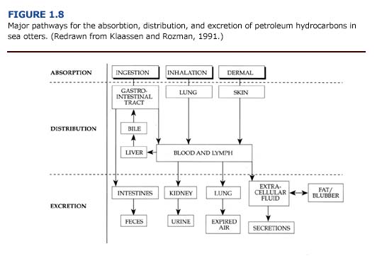
The absorption, distribution, and excretion of petroleum hydrocarbons involve many organ systems (Figure 1.8). Primary routes of exposure to petroleum hydrocarbons are ingestion, inhalation, and dermal absorption involving the gastrointestinal system, lungs, and skin, respectively. The liver and kidneys have a high capacity for binding toxicants and are considered major sites of toxicant concentration and accumulation (Klaassen and Rozman, 1991). Enzymes for the biotransformation of toxicants are located in these organs as well as the lungs and intestine (Sipes and Gandolfi, 1991). Petroleum hydrocarbons may be excreted through feces, urine, expired air, and, to a lesser extent, other secretions. Alternatively, petroleum hydrocarbons may be stored, and hence accumulate, in lipid rich tissues such as fat or blubber.

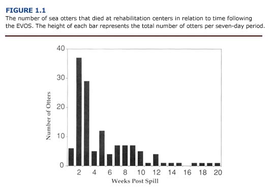
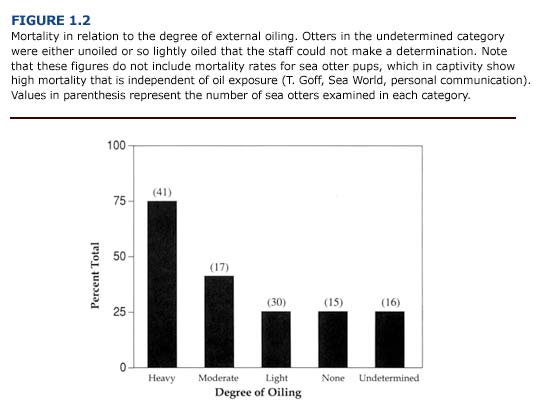
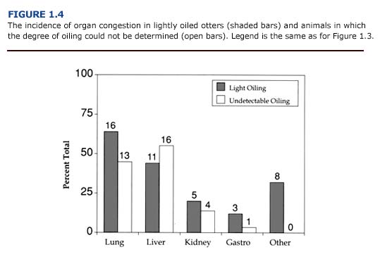
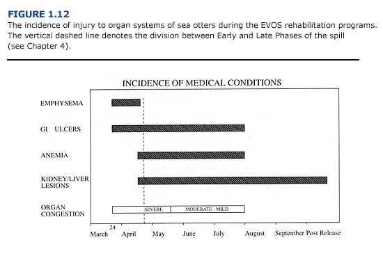
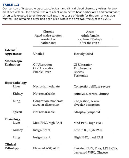
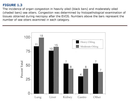
Macroscopic and microscopic evidence suggests that in contaminated sea otters, the liver, kidneys, gastrointestinal tract, and lungs are particularly vulnerable to oil, damage. Of the organ systems examined, the lungs had the highest incidence of lesions. Nearly 70% of the otters that arrived at rehabilitation centers during the first two weeks of the spill showed evidence of pulmonary damage (Williams et al., 1990). Other gross lesions included: 1) liver abnormalities in 55% of adult and juvenile otters, 2) gastrointestinal lesions in more than 50% of the otters, 3) cardiac lesions in 42% of the animals, and 4) kidney and spleen abnormalities in approximately 20% of the sea otters. Similar patterns are described for oiled sea otters by Lipscomb et al. (1994) and Mulcahy and Ballachey (1994). Specific lesions observed in sea otters during the EVOS are described below for each major organ system.

Liver

(a) Necropsy Observations. Because of its central role in metabolism, detoxification, and excretion of foreign chemicals, the liver is more likely to be exposed to petroleum hydrocarbons and their metabolites than other organs. Livers from oiled otters had a high incidence of lesions. Gross abnormalities included abnormal color and texture, enlarged lobes with prominent rounded edges, and atrophy. Livers were often friable and pale yellowish or reddish-brown in color with pale green mottling. There was no correlation between the degree of external oiling and the incidence of these lesions. Almost 75% of the unoiled animals examined had liver congestion, hemorrhage, necrosis, friable texture, or discoloration.
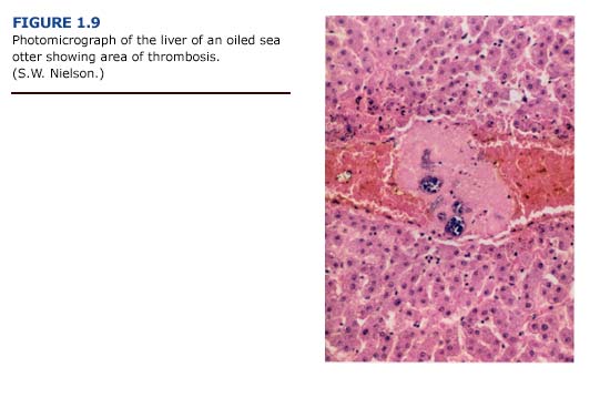
(b) Histopathology. Lesions of the liver were more impressive histologically than grossly. The histologic changes included congestion, hemorrhage, and a variety of degenerative changes such as hydropic degeneration, fat accumulation, and centrilobular necrosis. Necrosis was occasionally severe, with confluence of adjacent lobules to form larger areas. Generally, the centrilobular areas showed the most damage (Figure 1.9, see plate facing page 12). Periportal to diffuse hepatic lipidosis has been reported previously in oiled sea otters from the EVOS (Lipscomb et al., 1993, 1994).
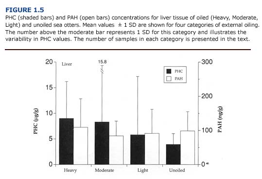
(c) Toxicology. Of the tissues examined, the liver generally contained the highest concentrations of PHC and PAH (PHC range = 0-36.4 ug/ g; PAH range = 0-234.9 ng/g; n = 21 oiled and unoiled animals). The levels of petroleum hydrocarbons in the liver did not correlate with the severity of external oiling or histologic lesions (Figure 1.5). For example, the liver from one heavily oiled animal with high concentrations of PAH and PHC appeared normal, with the exception of minor discoloration. Histopathological findings for this animal were also minor and consisted of periportal fatty change. In contrast, marked- to-severe congestion and necrosis of the liver was often accompanied by undetectable-to-moderate accumulation of petroleum hydrocarbons in other oiled and unoiled animals.
(d) Clinical Pathology. Blood chemistry panels indicated hepatic dysfunction for many of the oiled otters that died (Appendix 3, Figure E Download PDF). Plasma concentrations of alanine aminotransferase (ALT) and aspartate aminotransferase (AST) were elevated in these animals. Also, oiled otters were often hypoglycemic, although this may have been caused by many factors. (See Chapter 5.)
(e) Summary. The pattern of liver necrosis and congestion in oiled otters is compatible with both cardiovascular and toxicological damage. Cellular damage was often located in centrilobular rather than periportal areas (Figure 1.9). We found thrombi formation leading to vessel occlusion and local congestion in the livers. As a result, the centrilobular hepatocytes, which are located furthest from oxygen- carrying blood vessels, were the most likely to become hypoxic and necrotic. This pattern was evident in the majority of sea otters regardless of the degree of external oiling or level of petroleum hydrocarbon accumulation. Shock and hypothermia are the probable causes of the cardiovascular insufficiency which led to these patterns of liver damage. These findings suggest that liver damage and dysfunction in oiled sea otters results primarily from vascular congestion with systemic petroleum hydrocarbon toxicity playing a secondary role.


Kidneys

(a) Necropsy Observations. Macroscopic examination of the kidneys revealed few abnormalities in oiled and unoiled sea otters. Cortical streaking, pale coloration, and mild congestion were occasionally noted, but these did not correlate with the degree of external oiling or histologic findings.
(b) Histopathology. In contrast to the macroscopic findings, the microscopic renal lesions correlated well with the degree of external oiling. Kidneys from heavily and moderately oiled otters typically showed moderate to severe levels of congestion (Figure 1.3). Severe to moderate tubular lipidosis was commonly observed in these animals (present study, Lipscomb et al., 1993, 1994). Fewer remarkable lesions were observed in otters with less severe degrees of oiling.
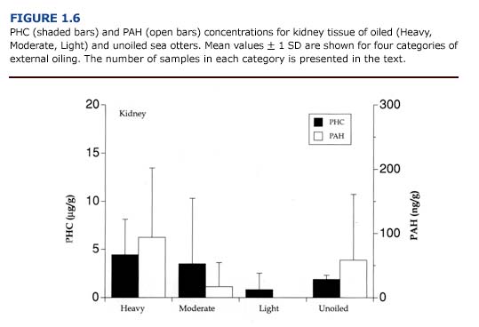
(c) Toxicology. PHC and PAH levels in the kidneys (Figure 1.6) were generally lower than reported for the liver (Figure 1.5). As observed for the liver, there was no apparent correlation between the concentration of petroleum hydrocarbons in the kidneys and the degree of external oiling. Likewise, the level of petroleum hydrocarbons did not always correspond to histopathological findings. Petroleum hydrocarbons appear to be cleared rapidly from the sea otter kidney. Samples of kidney from oiled otters that survived at least eleven days showed no evidence of petroleum hydrocarbon accumulation regardless of the degree of external oiling.
(d) Clinical Pathology. Acute renal insufficiency was evident for oiled sea otters and was manifested as elevations in serum concentrations of blood urea nitrogen (BUN), phosphorus (Appendix 3, Figures C, D Download PDF ) and creatinine. A complete description of this condition is provided in Chapter 5.
(e) Summary. Despite the importance of the kidneys as a site for biotransformation and excretion of toxicants, there was little accumulation of PAH or PHC in this tissue. Renal lipidosis was found only in otters that had hepatic lipidosis (Lipscomb et al., 1993). Many factors including infection, petroleum hydrocarbon exposure, mobilization of fat reserves during periods of reduced caloric intake, and hypoxia induced by vascular collapse may have contributed to this condition.

The incidence of organ congestion in heavily oiled and moderately oiled sea otters.

Lungs

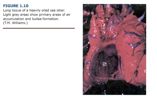
(a) Necropsy Observations. One of the most common lesions observed in otters captured during the first three weeks of the EVOS was bullous interstitial emphysema, which was characterized by the accumulation of interlobular air bubbles. In its most severe form, air was trapped along the trachea, mediastinum, and subcutaneous areas of the thorax. Emphysematous areas were patchy in distribution with no apparent preferential site. They ranged in size from a few millimeters to bullae formation of several centimeters in diameter (Figure 1.10, see plate facing page 12). The lungs typically appeared normal in color. Cases of subcutaneous emphysema were generally present only in animals with the most severe bullae formation in the lungs. The incidence and severity of emphysema generally correlated with the level of external oiling. Heavily oiled adult animals showed gross or histologic evidence of moderate to severe emphysema, usually involving interstitial tissues. In contrast, only mild forms of emphysema were noted for thirteen lightly oiled adult otters. Emphysema was also observed in one otter that was apparently unoiled but had sustained traumatic injuries during a fall. Depending on the severity, subcutaneous emphysema was detectable by palpation and correlated with macroscopic lesions observed during necropsy.
(b) Histopathology. Although histopathological examination of lung tissues demonstrated focal areas of congestion, edema, and alveolar distension, there was little indication of alveolar emphysema characteristic of chronic emphysema in humans. The histologic alveolar changes were fairly mild and subtle despite the incidence of severe interstitial and subpleural emphysema with bullae formation. There was no morphological evidence of damage to the airways or alveolar walls. Subpleural, interstitial, or subcutaneous bullous emphysema, rather than pulmonary or alveolar emphysema per se, are more appropriate terms for the conditions observed in oiled sea otters.

Pneumonia induced by the aspiration of oil, a characteristic respiratory lesion of petrochemical poisonings (Hatch, 1988), was not apparent in oiled sea otters during the EVOS.


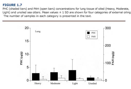
(c) Toxicology. The levels of PAH and PHC were comparatively low in the lungs (Figure 1.7). Mean PHC levels in oiled otters were 31-68% of the mean levels for liver. PAH concentrations were less than 20% of the values for liver samples. Unlike the kidneys and liver, there was a good correlation between petroleum hydrocarbon accumulation and tissue injury (i.e. emphysema). The average hydrocarbon concentrations in the lungs of otters showing severe congestion or emphysema were 3.68 %B1 3.19 ug/g for PHC and 21.14 %B1 14.01 ng/g for PAH (n = 5). In animals with mild or moderate congestion or emphysema, PHC levels were 1.42 %B1 1.00 ug/ g and PAH concentrations were 4.13 %B1 5.45 ng/ g (n = 9). Likewise, circulating levels of paraffinic hydrocarbons correlated well with the severity of emphysema (Chapter 4).
(d) Clinical Pathology. None of the standard tests for hematology or blood chemistry provided insight into lung damage. In the future, the partial pressures and concentrations of blood gases may prove useful in assessing the severity of respiratory injury in oiled sea otters.

(e) Summary. The pathogenesis of emphysema in oiled otters is not understood. Contributing factors may include weakening of the respiratory mucosa by petroleum hydrocarbon vapors and mechanical damage associated with respiratory exertion during capture, diving, coughing or agonal death. Damage associated with exposure to oil is supported in part by the positive correlation between blood and tissue petroleum hydrocarbon concentrations and the severity of emphysema. Because the incidence of emphysema was greatest during the first three weeks of the spill, exposure to the volatile aromatic hydrocarbons in fresh crude oil may have contributed to the development of the lesions. Twenty-five of forty-one cases of emphysema occurred within fourteen days of the EVOS. Only two instances of severe bullous interstitial emphysema were recorded after the first three weeks of the spill. Histopathological findings provide little insight into the pathogenesis of the lung lesions. Bullous emphysema has not been previously reported as a major injury in oiled marine mammals, except for a brief reference to the condition in one of four experimentally oiled polar bears (Oritsland et al., 1981).

Ventilatory exertion has been implicated in the development of spontaneous subcutaneous cervical and mediastinal emphysema in humans (Parker et al., 1990). Similarly, altered respiratory mechanics may also contribute to respiratory injury in oiled sea otters. Irritation of bronchial airways by volatile petroleum hydrocarbons may cause bronchial constriction. Extraordinary ventilatory exertion during such spasms could result in pulmonary distension and induce bullae formation. Predive inspiration, exercise-induced hyperventilation, extraordinary inspiratory effort due to blocked nares, and agonal breathing at death also may cause airway distension. Most likely, the cause of emphysema in oiled sea otters is a combination of chemical factors such as exposure to fresh or irritating crude oil, and mechanical factors including forced ventilatory expiration coupled with bronchial constriction.

Gastrointestinal System

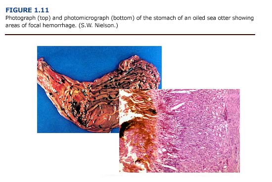
(a) Necropsy Observations. Gastrointestinal lesions were observed in sea otters throughout the rehabilitation process. The most frequently observed gastrointestinal lesions were areas of focal hemorrhage, ulceration, inflammation, and parasitism (Figure 1.11, see plates facing page 13). Gastrointestinal mucosal ulcerations, primarily involving the pylorus and the proximal small intestine, were a common finding. The consistency of the gastric and intestinal contents varied from a watery to a viscous dark brownish-green to reddish-black fluid. Several animals had dark black, tarry intestinal contents, although tests were not conducted to confirm the presence of oil or blood in the feces. In a related study, Mulcahy and Ballachy (1993, 1994) reported petroleum hydrocarbon levels indicative of oil ingestion in three of ten otters examined from the EVOS. Intestinal parasitism, primarily due to acanthocephalans, was a common finding. Cestode and nematode infestation frequently occurred. Ulceration of the oropharyngeal mucosa, especially the gingival mucosa, was observed, and intestinal intussusception occurred in several animals.
(b) Histopathology. Because of the focal nature of these lesions, histopathologic observations did not always correspond to macroscopic reports. Thus, a higher incidence of ulceration was reported during postmortem examinations than from the histopathology. Gastrointestinal lesions were found in both unoiled and oiled otters. The gastric mucosa showed acute focal changes; these ranged from necrosis and ulceration to acute hemorrhaging with free hemolyzed blood in the intestinal lumen.

(c) Toxicology. No toxicological tests were conducted on tissues from the gastrointestinal tract. In the future, petroleum hydrocarbon analyses of stomach contents and bile could prove valuable by providing information about the ingestion and excretion of crude oil.

(d) Clinical Pathology. Anemia may have been a secondary effect of gastrointestinal hemorrhaging. This is discussed in Chapter 5.
(e) Summary. It is difficult to relate the degree of external oil contamination to gastrointestinal abnormalities in oiled sea otters. There was a positive correlation between the degree of oiling and the incidence of stomach ulcers, but not for intestinal ulcers (Williams and Davis, 1990). Gastrointestinal hemorrhaging and superficial ulceration of the stomach mucosa may have been caused by shock, stress, or hypothermia (Paton, 1991). Stress induced gastrointestinal hemorrhaging is common in many mustelid species, including otters. In tests with laboratory animals, many components of crude oils are not considered ulcerogenic (Beck et al., 1984). Therefore, it is unlikely that petroleum hydrocarbon exposure alone would cause the degree of gastric erosion observed. Because lesions were evident throughout the rehabilitation process, stress, hypothermia, shock, parasite infestation, and oil contamination are all considered contributing factors of gastrointestinal lesions in oiled sea otters.

Photograph and Photomicrograph of the stomach of an oiled sea otter

Other Organs and Tissues

Adrenal hyperplasia, immunosuppression, and anemia have been identified as potential problems for oiled wildlife placed in rehabilitation centers (Leighton, 1991; White, 1991). The cause of these problems has been attributed to the combined effects of stress and oil toxicosis.

Adrenal hyperplasia, one manifestation of chronic stress, could not be confirmed macroscopically or microscopically in sea otters. Although enlarged adrenals were frequently noted at necropsy, that they were not weighed prevented a quantitative comparison with normal glands. However, hyperplasia of adrenal tissue was noted histologically for six of the animals examined. The incidence of hyperplasia did not correlate with the duration of captivity or degree of oiling. Histological examination showed that 60% of the otters (n = 32 animals) had no remarkable adrenal lesions.

Immunosuppression may be caused by a variety of petrochemicals. The polycyclic aromatic hydrocarbons (PAHs) in particular may produce a marked and prolonged depression in both primary and secondary serum antibody responses (Dean and Murray, 1991). Indirect evidence of immunosuppression has been observed for oiled polar bears (Oritsland et al., 1981) and oiled sea otters (Williams and Davis, 1990). Indications included abnormal inflammatory responses to sterile injections and to routine cuts and abrasions. Lesions were observed in the lymph nodes of approximately 45% of the heavily and moderately oiled sea otters. Moderate atrophy, histiocytosis, congestion, and hemorrhaging of lymph tissues were reported. The mechanisms by which immunosuppression develop are numerous and difficult to assess based on hematology; white blood cell counts were highly variable for all sea otters. Detailed examination of the cellular components of the bone marrow were not conducted. Although immunosuppression may occur in oiled sea otters, further research is needed to determine the incidence and the severity of the problem. Many factors, including exposure to petroleum hydrocarbons, hypothermia, stress, nutritional changes, and captivity, may challenge the immune systems of oiled animals during rehabilitation.

Anemia occurred frequently. Heavily and moderately oiled otters showed a higher incidence of anemia than lightly oiled animals. In general, the packed cell volume (PCV) of newly captured otters was within the normal range of 40-66% for healthy adult sea otters (Thomas Williams, in press); the values did not correlate with the degree of oiling or survivorship. Decreases in PCV occurred after several weeks in captivity, with a return to normal levels within four months (Williams, 1990). (See Chapter 5 for a discussion on anemia in oiled sea otters.)

ch1-clinical

Clinical, Toxicological, and Histopathological Profiles of Oiled Sea Otters

There is no typical clinical, macroscopic, and microscopic profile for oiled sea otters. Not all otters will exhibit the same spectrum of lesions in all organs susceptible to damage. However, there are several noteworthy trends concerning the incidence of tissue injury for sea otters exposed to oil (Figure 1.12). The incidence of each condition will vary with: 1) type of oil and degree of weathering, 2) duration and extent of exposure, 3) age, sex, and reproductive status of the animal, 4) nutritional state and health of the animal before contamination, and 5) environmental conditions and stressors (temperature, captivity, etc.).

THe incidence of injury to organ system of sea otters during the EVOS rehabilitation programs

Interstitial and subcutaneous emphysema will usually occur during the first weeks of a spill, when the greatest concentrations of volatile hydrocarbons are present. If a heavily or moderately oiled otter be- comes hypothermic, vascular congestion can occur at this time in many organs. The effects of vascular congestion on organ function may persist long after the hypothermic event. The resulting liver and kidney dysfunction, as indicated by serum chemistry, may persist for several months. Although not usually evident upon capture, anemia may develop after the first week of captivity and persist for several months. Gastrointestinal hemorrhaging can occur throughout the rehabilitation process as a result of many factors, including oil ingestion, hypothermia, parasite infestation, and stress.

Chronic and acute exposure to oil will also result in different pathologic profiles. Table 1.3 compares the macroscopic, microscopic, and toxicologic evaluations for an aged otter that resided in an active boat harbor in Alaska to those of an adult otter that was heavily oiled during the EVOS. Both animals showed lung congestion and distension, gastrointestinal ulceration, and hepatic lesions. There was evidence of petroleum hydrocarbon accumulation in the livers of both otters. In addition to these findings, the acutely oiled otter showed an accumulation of petroleum hydrocarbons in the lungs and the kidney. Emphysema and lesions within the lymph nodes were also apparent in this animal. While the harbor animal succumbed to the cumulative effects of organ failure associated with old age, the heavily oiled otter died within two weeks of the EVOS.

Comparison of histopathologic, toxicological and clinical blood values for two adult sea otters

ch1-summary

Summary

Our experience during the EVOS demonstrated the value of performing macroscopic and microscopic examinations on oiled sea otters that died. It also indicated the importance of baseline measurements for wildlife before an oil spill. Detailed postmortem examinations, histopathologic assessment, and blood chemistries provide the most valuable information for veterinarians caring for sea otters in the rehabilitation centers. Conversely, toxicological tests are comparatively expensive, time consuming, and often yield ambiguous results.

Major factors contributing to the mortality of oiled sea otters appear to be: 1) hypothermia, 2) shock and secondary organ dysfunction, 3) interstitial emphysema, 4) gastrointestinal ulceration, and 5) stress during captivity. Direct oil toxicosis may be a contributing factor, but is difficult to verify in otters contaminated during an oil spill.