ch4-lit

Literature Cited

Costa, D. P.1982. Energy, nitrogen, and electrolyte flux and seawater drink- ing in the sea otter, Enhydra lutris. Physiological Zoology 55 (1): 35-44.

Geraci, J. R., and V. J. Lounsbury. 1993. Marine mammals ashore: A field guide for strandings. Galveston: Texas A%26M Sea Grant.

Klaassen, C. D., and D. L. Eaton. 1991. “Principles of toxicology.” In Toxicology: The basic science of poisons. M. O. Amdur, J. Doull, and C. D. Klaassen, eds., 12-49. New York: Pergamon Press.

Mulcahy, D., and B. Ballachey. 1993. “Hydrocarbon concentrations in tissues of sea otters collected following the Exxon Valdez oil spill.” In Abstracts of the Exxon Valdez oil spill symposium. Anchorage, Alaska, Feb. 2-5, 1993: 293- 295.

Neff, J. M. 1990. “Composition and fate of petroleum and spill-treating agents in the marine environment.” In Sea Mammals and oil: Confronting the risks. J. R. Geraci and D. J. St. Aubin, eds., 1-33. San Diego: Academic Press, Inc.

St. Aubin, D. J. 1990. “Physiologic and toxic effects on polar bears.” In Sea mammals and oil: Confronting the risks. J. R. Geraci and D. J. St. Aubin, eds., 235-39. San Diego: Academic Press, Inc.
Williams, T. M. 1990. Evaluating the long term effects of crude oil exposure in sea otters. Wildlife Journal 13 (3): 42-48.

Williams, T. M., and R. W. Davis. 1990. Sea otter rehabilitation program: 1989 Exxon Valdez oil spill. Report to Exxon Company, USA. International Wild- life Research.

Williams, T.M., R. A. Kastelein, R. W. Davis, and J. A. Thomas. 1988. The effects of oil contamination and cleaning on sea otters (Enhydra lutris). I. Thermoregulatory implications based on pelt studies. Canadian Journal of Zoology 66 (12): 2776-81.

Williams, T.M., J. McBain, R.K. Wilson, and R.W. Davis. 1990.”Clinical evaluation and cleaning of sea otters affected by the T/V Exxon Valdez oil spill”. In Sea otter symposium: Proceedings of a symposium to evaluate the response effort on behalf of sea otters after the T/V Exxon Valedz oil spill into Prince William Sound, Anchorage Alaska, 17-19 April 1990. K Bayha and J. Kormendy, eds. U.S. Fish and Wildlife Service Biological Report 90 (12): 236-57.

ch5-intro

Chapter 5 – Introduction

The clinical disorders exhibited by oiled sea otters will depend on the type of oil encountered and the degree and duration of exposure. Unfortunately, much of this information is unavailable when otters are captured during an oil spill. The route of exposure and duration of contamination can only be inferred from the date of the spill, the rate of oil weathering, and the movement of the oil into otter habitats. To overcome this problem, Williams et al. in Chapter 4 suggest that oil spills be divided into Early and Late Phases. This division enables veterinarians to plan for the type of clinical problems most often encountered when the oil is concentrated and fresh, or after the oil has dispersed and weathered.

Depending on the rate of weathering, the detrimental effects of oil are greatest during the first two to three weeks or Early Phase of a spill (Neff, 1990). During this period, otters often arrive at the rehabilitation center completely covered with fresh oil and displaying the severest medical problems. These animals require immediate and often long-term care if they are to survive. In contrast, the period of treatment and recovery may be short for otters lightly contaminated with weathered oil during the Late Phase of a spill. Some Late Phase otters may even be healthy enough to bypass the rehabilitation process and be sent directly to a prerelease facility. It is important to remember that the primary objective of any clinical regimen will be the graduation of otters through the successive stages of rehabilitation for the purpose of release.

This chapter describes the etiology, clinical manifestations, and treatment of specific disorders that commonly occur in oiled sea otters. A summary of symptoms and recommended treatments is provided in Table 5.5 beginning on page 84. We divide the chapter into three sections: disorders that occur during the Early Phase of a spill, disorders common to both phases of an oil spill, and long-term treatments. Veterinary clinicians are also referred to Chapter 1 for a discussion of the pathology, toxicology, and clinical history of oiled sea otters during the Exxon Valdez oil spill (EVOS) and to Chapter 4 for emergency treatment methods and assessment of oil exposure. Appendix 6 Download PDF lists the equipment required for treating oiled sea otters.

ch5-disorders-early

Disorders Common to the Early Phase of Oil Spills

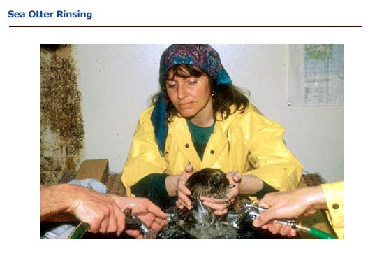
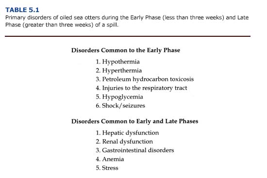
Whether the cause is related to oil exposure or stress, six primary medical problems have been identified for Early Phase sea otters (Table 5.1). The majority of sea otters arriving at the rehabilitation center during the Early Phase will be heavily or moderately oiled and require immediate cleaning. Special care should be taken when handling animals exhibiting signs of hypothermia or respiratory distress. Unnecessary movement or agitation may induce cardiac arrhythmias and interstitial emphysema associated with these conditions (see below).

Primary disorders of oiled sea otters during the Early Phase and Late Phase of spill

Hypothermia

(a) Etiology. For most mammals, hypothermia is defined as a core temperature less than 35°C (95°F) (Knochel, 1985). As body temperature declines, heat production is increased by shivering and heat loss is reduced by decreased peripheral blood flow. If the core temperature drops below 32°C (90°F), shivering ceases, muscle tone increases, and the animal may appear in rigor mortis.

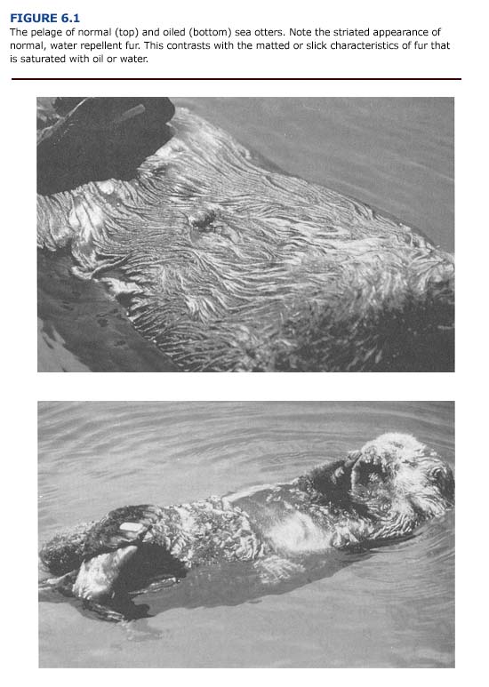
Hypothermia is a serious threat to sea otters during an oil spill. Because oil destroys the insulating quality of the otter’s fur, contamination can result in a rapid decrease of core temperature, especially if the animal remains in the water or is exposed to wind, rain, and cold air temperatures. Oiled otters often forgo feeding to haul out on shore or spend additional time grooming their contaminated fur. The result is a rapid decline in food intake, which can result in hypoglycemia and dehydration, factors that further predispose the otter to hypothermia.

(b) Clinical Manifestations and Diagnosis. The normal rectal temperature of sea otters ranges from 37-39°C (98.6-102°F). During the EVOS, more than 36% of heavily and moderately oiled sea otters arriving at rehabilitation centers were diagnosed as hypothermic. The lowest core body temperature recorded was 29.4°C (85°F) for an otter that arrived cyanotic and unconscious.

Clinical manifestations of hypothermia include locomotor incoordination, disorientation, and lethargy. Peripheral vasoconstriction and shivering are frequent physiological manifestations of mild hypothermia as core temperature declines to 32°C (90°F). At lower core temperatures, hyporeflexia, stupor, cessation of shivering, and muscle rigidity become evident (Knochel, 1985). Left untreated, the hypothermic animal will become unconscious. Reductions in heart rate, blood pressure, peripheral vascular resistance, cardiac output, and central venous pressure occur during severe hypothermia. These cardiovascular changes have a profound effect on organ function and may lead to long-term cellular damage, particularly in metabolically active tissues such as the liver and brain (see Chapter 1). In view of this, the attending veterinarian must consider the possibility of a previous hypothermic event and consequent organ damage for oiled otters, despite the presentation of a normal body temperature during initial examinations.

One of the greatest dangers associated with hypothermia is cardiac arrhythmias, which can result in ventricular fibrillation and death, particularly at core temperatures below 28°C (82°F) (Knochel, 1985). Severe shivering contributes to lactic acid accumulation and resultant metabolic abnormalities. Metabolic acidosis and hyperkalemia may occur if hypothermia is prolonged. The concomitant metabolic imbalance leads to cardiac arrhythmias (Bowen and Bellamy, 1988). Atrial fibrillation and ventricular tachycardia also may occur in cases of severe hypothermia. Physical stimulation predisposes the animal to the development of these arrhythmias. Therefore, handling and physical restraint of the hypothermic otter should be minimized.

Creatine phosphokinase (CPK) increases in the blood during severe hypothermia as a result of cellular damage. We found that serum CPK was elevated in 68% of the oiled otters that died during the EVOS (Appendix 3, Figure F Download PDF). However, CPK also may increase from handling stress and from cardiac and skeletal muscle damage (capture myopathy syndrome) not associated with a hypothermic event (Bossart and Dierauf, 1990). Therefore, CPK should not be considered a diagnostic indicator of cellular damage resulting exclusively from hypothermia.

(c) Treatment. We recommend measuring the core temperature of all sea otters entering the rehabilitation center. Body temperature should also be measured every thirty minutes in anesthetized otters during cleaning. A digital thermometer with a flexible thermocouple probe (Physiotemp, Inc.) should be used. The probe should be inserted at least fifteen cm into the rectum and may be left in place during cleaning and treatment. Glass thermometers are not recommended.

Treatment of the hypothermic animal involves internal and external rewarming and will depend on the state of consciousness and degree of oiling. Passive rewarming at a rate of 0.5°C (1°F) per hour is optimal (Knochel, 1985). Often the core temperature of mildly hypothermic otters will return to normal without additional rewarming therapy the animal is placed in a warm (20°C; 68°F) room. Alert animals may facilitate rewarming by grooming or shivering. The animals should be placed in a dry, well-ventilated cage during this period. If cleaning is delayed, the coat of the otter should be dried with towels or a pet dryer set at room temperature to reduce further heat loss.

Usually, the hypothermic otter is extensively covered with fresh crude oil and is lethargic. In cases of severe hypothermia (core temperature less than 32°C or 90°F) or prolonged hypothermia lasting more than twelve hours, active rewarming is recommended (Zenobl 1980). Laying the hypothermic otter on a recirculating warm water veterinary pad (Aquamatic K Pad, American Hospital Supply) or plastic bags filled with warm water will enhance rewarming. Sedated or lethargic otters thermoregulate poorly and will rapidly gain or lose heat during cleaning (Davis et al., 1988). The veterinarian may use this as an opportunity to slowly rewarm the hypothermic otter by maintaining the wash and rinse water temperatures between 37-40%B0C (98.6-104°F).

Active external rewarming by immersion in warm water is potentially dangerous. External rewarming may cause rewarming shock when lactic acid washed out of previously hypoxic tissues leads severe metabolic acidosis (Knochel, 1985). Earlier studies also describe a paradoxical decrease (after-drop) in core temperature due to peripheral vasodilation associated with warm water immersion in chronic cold-stress patients. It was believed that cold blood returning from the periphery cooled the myocardium and increased the likelihood ventricular fibrillation. However, in more recent investigations, the phenomenon of after-drop in body temperature has been difficult to document. There are insufficient stores of blood in vasoconstricted peripheral areas to cause a decrease in myocardial temperature (Lloyd 1986). Rather than an after-drop in core temperature, the balance between the size of the vascular bed and the circulating blood volume (both of which depend on vasomotor tone and state of hydration) was identified as a critical factor for the survival of humans during rewarming. Rewarming by warm water immersion is currently considered beneficial if cardiovascular and respiratory functions a monitored. Thus, there is growing belief that rewarming by warm water immersion is a fast, effective way of treating hypothermia when the patient is closely monitored. This method is recommended for severely hypothermic animals when core temperature is below 32°C ( 90°F). Water temperature should be 37-40°C (98.6-104°F) and the animal’s vital signs, especially heart rate, must be monitored throughout rewarming.

Several methods of internal rewarming are possible (e.g. high color irrigation, peritoneal dialysis, hemodialysis, intragastric lavage). Most of these techniques are impractical in rehabilitation centers or may cause additional medical complications for animals that are already severely stressed. Rewarming by the administration of warm fluids the safest and preferred method of treatment. Fluid replacement provides additional benefits by improving peripheral circulation and the cardiac output of a hypothermic animal. The fluids should be prewarmed to 37-39°C (98.6-102°F) by passage though a hot water bath or by a bacteriologic incubator. The fluids may be administered subcutaneously or intravenously through the jugular vein or popliteal vein. If the animal is unconscious or sedated, large bore jugular catheters can be used effectively. Lactate-free and potassium-free fluids such as normal saline or a 1-to-1 mixture of 5% dextrose and normal saline (20 ml/kg SQ or IV) are preferred because of the electrolyte and metabolite imbalance of hypothermic patients. A solution containing 10-20% dextrose (10-20 ml/kg IV) is recommended for otters that are hypoglycemic as well as hypothermic. Plasma pH and electrolyte concentrations should be monitored hourly until core temperature returns to 37-39 (98.6-102°F).

During rewarming, sodium-potassium exchange accelerates. As a result, hypokalemia may occur, which can cause cardiac arrhythmias. If cardiac failure occurs, the heart of a hypothermic animal may be unresponsive to lidocaine injections. Cardiopulmonary resuscitation (CPR) and the administration of oxygen should be initiated and continued while core temperature is being raised (Zenoble, 1980). CPR, oxygen administration, intravenous glucose, and warm water immersion were effective in reviving an unconscious, severely hypothermic otter during the EVOS.

Complications following rewarming can include pneumonia, gastric erosions, intravascular erosions, and acute renal tubular necrosis, with pneumonitis the most common problem in human patients (Bowen and Bellamy, 1988). Antibiotic therapy following rewarming along with corticosteroids to combat shock are recommended. Note that the delayed metabolism of drugs in hypothermic animals predisposes them to over medication.

In summary, all animals should be monitored closely during rewarming procedures. Passive rewarming at a rate of 0.5°C (1F) per hour in a room at 20°C (68°F) is the preferred treatment for mildly or moderately hypothermic otters. Severe hypothermia (core temperature less than 32°C or 90°F) should be treated by intravenous or subcutaneous administration of warm, normal saline or a 1-to-1 mixture of normal saline and 5% dextrose. External rewarming by immersion in warm water also is recommended, but the veterinarian or animal care specialist must closely monitor the animal’s vital signs.

Hyperthermia

Panting, dry mucous membranes, lethargy, hind flippers that are warm to the touch, and a core temperature exceeding 39°C (102°F) are manifestations of hyperthermia in sea otters. This condition can occur during transport, anesthesia, or whenever caged otters are placed in a poorly ventilated area warmer than 20°C (68°F) without access to water or ice. Despite the decrease in insulation resulting from oily fur, sea otters easily overheat when out of water. Excessive grooming, inadequate ventilation in transport cages, and hyperactivity associated with handling exacerbate the problem.

Hyperthermia in sea otters is easily prevented by placing the animals in seawater at normal seasonal ocean temperatures. Otters in dry cages should be kept in well ventilated areas at temperatures near 15°C (60°F). The grated bottom of transport cages or critical care cage should be partially covered with chipped ice to enable the otter to cool itself. The otter may also eat the ice which provides additional cooling and helps to prevent dehydration. In extreme cases, where the animal’s core temperature exceeds 40°C (104°F), the hind flipper should be sprayed with water and packed in ice for short periods Because the flippers are well vascularized, cooling them will provide an immediate, short-term benefit to the overheated otter. Care should be taken to avoid localized vasoconstriction of the flippers due to prolonged contact with ice packs.

Petroleum Hydrocarbon Ingestion and Absorption

(a) Etiology. There is considerable confusion concerning the detrimental effects of petroleum hydrocarbon ingestion and absorption. Much of the confusion undoubtedly originates from the fact that oil is a complex mixture of aromatic and aliphatic petroleum hydrocarbons and inorganic compounds, each varying in toxicity. The situation is further complicated by the fact that the chemical composition of oil, and hence its toxicity, changes as it weathers and dissipates. Thus, marine mammals may be exposed to different concentrations of potentially harmful petroleum hydrocarbons during the course of a spill.

Each oil spill will be different, and the effects on wildlife will depend on the type of petroleum hydrocarbons encountered, the degree of weathering, and the duration of exposure. As discussed in Chapter 4, the level of toxicity and the probability of systemic hydrocarbon exposure are greatest during the first weeks of a spill when the oil is fresh and the concentration of aromatic hydrocarbons is highest. In the case of chronic spills, as may occur at marine oil terminals and in harbors, the period of toxicity may be prolonged. Ambient air and water temperatures, weather conditions, and sea state will greatly affect the rate of oil weathering.

Individual petroleum hydrocarbons may be cardiotoxic, hemotoxic, neurotoxic, or hepatotoxic and may induce central nervous system depression (Amdur et al., 1991). As a group, the polycyclic aromatic hydrocarbons (PAHs) are the most toxic. The inhalation of high concentrations of petroleum hydrocarbon vapors can cause excitement, depression, unconsciousness, and death; ingestion can cause severe diarrhea, cardiovascular collapse and organ degeneration (Coppock et al., 1986). The effects of benzene, an aromatic compound commonly found in crude oil, have been examined in studies using laboratory mammals. Exposure to benzene may lead to dose-dependent changes in hematological parameters and lesions in the thymus, bone marrow, spleen, and testes (Ward et al., 1982).

Damage to individual organ systems may occur by direct exposure to petroleum hydrocarbons or secondarily from toxic, metabolic by- products. The lungs, kidneys, and liver are target organs for many toxicants (Klaassen and Rozman, 1991). Petroleum hydrocarbons of high vapor pressure are eliminated through the lungs. The lungs also may reduce hydrocarbons into secondarily toxic metabolites. Because the liver is the site of detoxification and elimination of many compounds, it too is considered vulnerable to the effects of petroleum hydrocarbon absorption. The kidneys are susceptible to damage by toxicants because they receive a large portion (approximately 20%) of the cardiac output, and because tubular secretion and reabsorption may concentrate toxicants within cells. The immune and hematopoietic systems also may be affected.

(b) Clinical Manifestations and Diagnosis. The clinical manifestations of petroleum hydrocarbon toxicosis are difficult to distinguish from other medical problems exhibited by oiled otters. Information about the type of oil and date of the spill will aid the veterinarian in estimating the maximum duration of exposure and the relative toxicity of the contaminant. The degree of external and internal contamination may be assessed by visual examination of the pelt and by blood tests, as described in Chapter 4. Many heavily contaminated otters will spontaneously pass cestodes and acanthocephalids in their feces, providing another indicator of internal oil exposure.

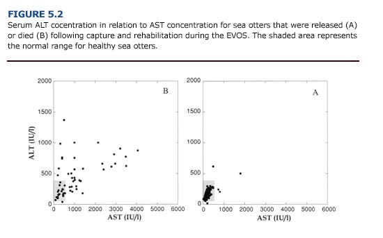
Otters exposed to petroleum hydrocarbons may appear normal during initial examination or display a range of clinical signs including excitability, seizures, CNS depression, lethargy, ataxia, emesis, diarrhea, respiratory distress, and cardiac arrhythmias. The detection of oil in the feces will confirm ingestion. A simple test is to suspend and shake fecal material in water; petroleum products will separate and float to the surface. Hepatocellular enzymes may be elevated (Appendix 3, Figure E Download PDF). Crude oil can be irritating to mucous membranes; corneal ulceration and photophobia may be apparent.

Aspiration pneumonia is a common and serious clinical disorder which can develop in cattle, cats, and dogs exposed to a variety of petroleum products (Hatch, 1988). However, this condition was never observed in sea otters during the EVOS.

(c) Treatment. To prevent further absorption or ingestion of crude oil, sea otters should be moved from the spill area and cleaned with detergent (see Chapter 6). Treatments should focus on delaying absorption and promoting the elimination of ingested oil. The induction of emesis for eliminating ingested petroleum compounds is not recommended due to its limited value when treatment is delayed more than two hours after oil ingestion and the high risk of inhalation pneumonia. Likewise, gastric lavage is not recommended. The oral administration of mineral oil (1 ml/kg) has been used to treat accidental kerosene poisoning in mammals, and may mitigate the absorption of ingested petroleum compounds (Bailey, 1980; Coppock et al., 1986). However, this technique has not been tried on oiled sea otters and it risks aspiration pneumonia associated with vomiting.

Adsorbents such as activated charcoal are often effective in reducing absorption of many ingested toxicants. Several products are available. ToxibanTM (6 ml kg; approximately 120 ml dose) was administered orally to heavily and moderately oiled sea otters during the EVOS. A slurry of activated charcoal and water is administered to sedated sea otters via a syringe connected to a stomach tube, (see section on hypoglycemia for details of tube placement). To reduce stress associated with handling and sedating the otter, we do not recommend a multiple treatment program. Other promising treatments include compounds, such as Questran TM, that bind bile acids in the gastrointestinal tract, thereby preventing hepatic recycling of toxicant: To date, this product has not been used on oiled sea otters.

Oral treatments are less effective against dermal absorption an inhalation of petroleum hydrocarbons. Absorbed compounds may b sequestered in fat or removed by the liver, kidneys, and lungs. The elimination process of some toxicants may be expedited by diuretic peritoneal dialysis, or manipulation of urinary pH (Bailey, 1980). However, we do not recommend these procedures for oiled sea otters. Immunosuppression, metabolic imbalance, impaired renal and hepatic function, and cardiac arrhythmias directly or indirectly associated wit the absorption of petroleum hydrocarbons may complicate these procedures. Most treatments for petroleum hydrocarbon exposure will be limited to supportive care and the mitigation of its effect on individual organ systems.

Injuries to the Respiratory Tract

(a) Etiology. Exposure of the respiratory tract to air borne or blood born petroleum hydrocarbons may lead to pulmonary damage and decreased gas exchange across the alveoli. The specific injury will depend on the route of exposure and can involve the upper and lower respiratory tract. Damage to the gas exchange surfaces will increase the work of breathing.

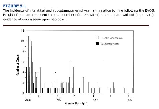
Injuries to the respiratory tract commonly occurred in oiled otters during the first three weeks of the EVOS. Over 75% of oiled sea otters brought to rehabilitation centers during this period showed respirator distress and interstitial emphysema (Figure 5.1; Williams and Davis 1990). The incidence of emphysema during the Early Phase suggests that exposure to volatile petroleum hydrocarbons was a significant contributing factor. Depending on environmental conditions, aromatic hydrocarbons (e.g. benzene, toluene, xylene) will evaporate within days of an oil spill. These are considered the most toxic compounds in crude oil and are known to cause damage to the lungs and mucous membranes of the bronchial airways (Geraci and St. Aubin, 1990).

The incidence of interstitial and subcutaneous emphysema in relation to time following the EVOS

(b) Clinical Manifestations and Diagnosis. The inhalation or aspiration of toxic compounds produces many pathologic changes in respirator tissues including: 1) bronchospasm, 2) impaired mucociliary clearance 3) mucosal sloughing, 4) atelectasis, and 5) pulmonary edema (Fm row, 1980). Tachypnea, congestion, and the use of accessory respiratory muscles for ventilation are typical clinical manifestations of a respiratory tract injury. The respiratory rate may be accelerated above twenty breaths/minute in oiled sea otters. In other mammals, long-term health problems following exposure to a variety of petroleum hydrocarbon can include aspiration and postinhalation pneumonia. However, his tologic examination indicated that pneumonia did not occur in sea otters that died during the EVOS (Chapter 1).

Respiratory tissue injury in oiled otters ranges in severity from irritation of the nasopharyngeal membranes and rhinitis to interstitial and subcutaneous emphysema. Rhinitis and sinusitis can be persistent problems in oiled sea otters. Clinical manifestations are epistaxis and purulent nasal discharge. During the EVOS, cultures and sensitivity tests revealed the presence of pathogenic E. coli and Proteus. Oral and pharyngeal cavities should be examined for edema and swelling. Discharges should be examined microscopically for cellular debris, red blood cells, neutrophils, and bacteria.

The emphysema observed in oiled sea otters may range in severity and location. The condition is classified as: 1) severe (bullous emphysema in the lungs and interstitial areas), 2) moderate (bullous emphysema throughout the lungs), 3) mild (focal areas of lung damage), and 4) none (no evidence of interstitial or subcutaneous emphysema). Subcutaneous emphysema is characterized by pockets of air below the skin. Small bubbles may be felt subcutaneously in the axillary region of the otters. In severe cases, air pockets can be felt or seen beneath the skin along both sides of the neck, thorax, and along the spine. Postmortem examination of otters that died with this condition during the EVOS revealed that the subcutaneous emphysema arose from ruptured membranes in the lungs. Air escaping from the lungs moved along the mediastinum, through the thoracic inlet and accumulated in subcutaneous tissues. To assess the presence of subcutaneous emphysema, the axillary and chest region of the animal should be palpated during the initial physical examination. Gas bullae may be felt as distortions below the skin and can be heard “popping” (crepitation) when pressed lightly. Roentgenographic examination can confirm this condition, but is of little practical use and subjects the animal to additional stress.

The development of interstitial and subcutaneous emphysema in oiled sea otters is not completely understood. One possible explanation is that chemical irritation of the airways from breathing petroleum hydrocarbon vapors leads to bronchial or laryngeal constriction. Nasal passages may become congested due to inflammation of the mucous membranes. When accompanied by labored breathing, alveolar rupture and bullae formation may ensue. Consequently, the handling of animals with suspected emphysema (i.e. heavily and moderately oiled otters captured during the first weeks of a spill) should be minimized (c) Treatment. The treatment of respiratory injuries is limited and based on methods for mitigating human injuries due to inhalation of toxic substances (Farrow, 1980). Further exposure should be prevented by removing the animal from the spill area and by cleaning contaminate fur. The correlation between the severity of emphysema and presence of volatile components of fresh crude (see Chapter 4) indicates that these measures should occur as soon as possible during the Early Phase of a spill. If the animal is agitated, diazepam (0.2 mg/kg PO or 0.1 mg/kg 1M) may be administered to prevent hyperventilation and to reduce the animal’s activity level and metabolic rate.

Rhinitis and sinusitis, as determined from cultures and sensitivity tests, should be treated with antibiotics. Initially the otters may be placed on emofloxacin (2.5 mg/kg bid IM or PO) for mature otter and amoxicillin (12 mg/kg bid IM) for immature otters. Although antibiotics may decrease the frequency of epistaxis and nasal discharge full recovery may take as long as three months. Respiratory parasites such as nasal mites should be controlled with Ivermectin (50 ug/kg 2 a single dose SQ or PO).

The treatment of interstitial and subcutaneous emphysema is limited to supportive care. In clinical settings, supplemental oxygen has been used to help alleviate respiratory distress. However, its large scale use for oiled sea otters is impractical. Positive pressure ventilation systems may aggravate this condition by inducing the formation (of bullae in alveolar membranes weakened by exposure to petroleum hydrocarbons; positive pressure delivery of oxygen is not recommended. Positive pressure and inhalation anesthetics during cleaning procedures also are contraindicated for otters showing signs of emphysema. Aminophylline (10 mg/kg bid PO, slow release form), bronchodilator, is recommended for any otter exhibiting respirator distress. However, aminophylline is a diuretic and can cause cardiac arrhythmias. Therefore, this treatment is not recommended for animals exhibiting renal insufficiency or hypothermia.

Hypoglycemia

(a) Etiology. Hypoglycemia (plasma glucose less than 60 mg/ dl) in oiled sea otters may result from: 1) inability to feed prior to capture and, reduction in glycogen stores, 2) fasting during capture and transpor1 3) impaired hepatic function or intestinal absorption, or 4) stress and shock. The high metabolic rate of sea otters in general (Costa and Kooyman, 1982), and oiled sea otters in particular (Davis et al., 1988) predisposes these animals to hypoglycemia when food is withheld for several hours. Other factors such as anorexia, hypothermia, and fever also may predispose them to hypoglycemia. Following the EVOS 40% (n = 27) of the otters that died were hypoglycemic (Appendix 3 Figure A Download PDF).

(b) Clinical Manifestations and Diagnosis. Symptoms of hypoglycemic include pulmonary edema, hypothermia, and central nervous system dysfunction (i.e. depression, seizures, muscular weakness, and locomotor incoordination). In severe cases, the animal may become unconscious, and prompt treatment is critical. Plasma glucose concentration should be measured during the initial physical examination of all oiled otters. Reagent strips (Gluco-StixTM, Ames Laboratories) provide a rapid, qualitative measure of blood glucose concentration. This allows the veterinarian to verify the hypoglycemia and initiate emergency treatments. We recommend a subsequent quantitative measurement of plasma glucose as soon as possible. Desktop blood chemistry analyzers (Eastman Kodak, Inc.; Abbot Laboratories) or small digital analyzers for routine blood glucose monitoring in diabetics provide rapid results from small quantities of blood.

(c) Treatment. Hypoglycemia is treated by increasing the otter’s intake of dextrose (glucose) as soon as possible. The voluntary ingestion of dextrose can be achieved by offering chipped ice balls containing a 50% dextrose solution. If the animal is lethargic or semiconscious, administer 10-20% dextrose (10-20 ml/kg IV to effect). The oral administration of 50% dextrose (1 ml/kg) through a stomach tube will provide an immediate but temporary increase in the plasma glucose concentration. In general, the otter will respond to treatment within thirty minutes of fluid administration, but relapses may occur if the underlying dietary, hepatic, or gastrointestinal problems are not resolved. Following initial treatment, hypoglycemic otters should be fed frequently (at least every hour) until stable. If seizures occur after blood glucose is stabilized, diazepam (0.1 mg/kg 1M) should be administered.

Insulin resistance may limit the effectiveness of prolonged glucose administration and carbohydrate feeding to supplement calories in animals that refuse to eat. If an otter can not be encouraged to eat voluntarily, enteral feeding (stomach tube feeding) may be necessary. Animals that will not eat utilize stored fat and tissue protein for metabolic energy. The primary objective of enteral feeding is to prevent the loss of tissue protein by providing nutrients that can be readily digested and absorbed.

During enteral feeding, the otter should be chemically restrained (see Chapter 3), although severely debilitated otters may be physically restrained by experienced handlers. A plastic or wooden dowel should be placed between the premolars, and a stomach tube inserted into the esophagus to a premeasured length (about twenty cm in adult otters). The tube can usually be seen or felt to pass on the left side of the trachea. Proper placement of the tube in the stomach should be tested by listening for bubbling sounds when air is blown into the tube. The enteral diet is injected into the feeding tube with a syringe. After feeding, the tube is sealed while it is withdrawn to prevent leakage of residual fluids into the pharynx.

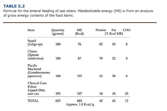
Enteral diets are slurries of common food items blended with a diluent of either liquid enteral (e.g. PedialyteTM) or water. Carnivores require more fat and protein than the enterals manufactured for humans. An example of a slurry suitable for sea otters is shown in Table 5.2. Resting adult sea otters require about 75 kcal/kg/ day in their diet in order to maintain their body weight (Davis et al., 1988); additional food energy would be required for an otter to gain weight. Because of stress caused by orogastric intubation, enteral meals are given only three times daily. Thus, meal size is relatively large. An otter weighing 40 kg requires 3.0 kg (1.0 kg per meal x 3) of the high-energy diet shown in Table 5.2 daily.

Formula for the enteral feeding of sea otters

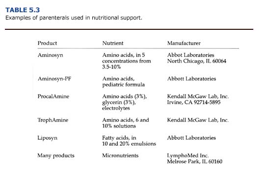
Calories and nutrients given parenterally consist of solutions of glucose, amino acids, and lipid emulsions administered intravenously (Table 5.3). For carnivores, dextrose (25 or 50% solutions), amino acids (3.5-10% solutions), and lipids (10 or 20% emulsions) are combined to provide about 35-45% of calories from carbohydrates, 20-25% of calories from amino acids, and 35-45% of calories from fat. Solutions of vitamins, trace minerals, and electrolytes should be provided as well. Parenteral nutrition is expensive, labor intensive, and risks sepsis. For sea otters, it should be used only for critically ill animals, and the switch to enteral or normal feeding should be made as soon as possible.

Examples of parenterals used in nutritional support

Shock

(a) Etiology. All forms of shock are characterized by acute circulatory insufficiency and inadequate capillary perfusion. Hypovolemic shock due to severe blood loss is rare in oiled sea otters, but should be considered in cases of gastrointestinal hemorrhage (see below). Oiled otters that are hypothermic or stressed may also experience shock due to reduced cardiac output or peripheral vasodilatation. A consequence of the reduction in tissue perfusion is localized hypoxic ischemia and impaired cellular function. To compensate for the general reduction in tissue circulation, the body may respond by readjusting blood flow to vital organs such as the heart and brain. At the cellular level, poor tissue perfusion will result in cellular swelling, intracellular acidosis (as a result of elevated lactic acid production), and extracellular acidemia. The onset of these conditions will depend on the animal’s body temperature.

(b) Clinical Manifestations and Diagnosis. It is critical that symptoms of shock be recognized and treated quickly. Primary signs of shock in otters are tachycardia, hyperventilation, depression, muscular weakness, cold hind flippers, and hypotension (poor capillary refill, reduced pulse pressure). Poor peripheral perfusion can be detected by the palpation of cold extremities and observing capillary refill times of mucous membranes. Refill times exceeding two seconds indicate inadequate peripheral perfusion. (c) Treatment. Treatments for shock will be specific for the individual otter and depend on underlying causes. Detailed treatments are complex and are described in detail elsewhere (see for example, Veterinary Pharmacology and Therapeutics, Iowa State University Press; Current Veterinary Therapy, W. B. Saunders Company). For most oiled sea otters, the veterinarian should initiate fluid volume expansion to reestablish adequate tissue perfusion as soon as possible. Administration of normal saline or a 1-to-1 mixture of normal saline and 5% dextrose (20 ml/kg IV) is recommended. Sodium bicarbonate (1 mEq/ kg IV or 50 mg/kg PO) or supplemental dextrose (1 ml/kg of a 50% dextrose solution by stomach tube) may be indicated, if the animal is acidotic or hypoglycemic, respectively. Finally, dexamethasone (1-2 mg/kg/ day 1M) or methylprednisolone (0.06 mg/kg / day 1M or IV) should be administered.

Seizures

(a) Etiology. Seizures in oiled sea otters may be caused by:
1) hypoglycemia,
2) hypothermia or hyperthermia,
3) hepatic encephalopathy,
4) electrolyte imbalances,
5) dehydration,
6) sepsis,
7) exposure to petroleum hydrocarbons, or
8) adverse reaction to anesthetics (i.e. fentanyl).
Periodic seizures in some animals may persist for weeks to months, slowly reducing in frequency and intensity over time.

(b) Clinical Manifestations and Diagnosis. Depending on the cause, seizures or convulsions are characterized by one or all of the following signs: unconsciousness, loss of (flaccid) or excess (clonus to rigidity) muscle tone, changes in the autonomic nervous system (urination, salivation, defecation, vomiting), and behavioral abnormalities (vocalization, pacing) (Parker, 1980).

(c) Treatment. As with shock, the treatment of seizures will depend on the underlying cause(s). For repeated or prolonged seizures, anticonvulsant drugs may be warranted (diazepam, 0.2 mg/kg PO or 0.1 mg/kg 1M). In cases of hypoglycemic seizures, anticonvulsants are not necessary; these animals will respond rapidly to glucose administration (see previous section on Hypoglycemia). Seizures associated with hepatoencephalopathy may be reduced by the oral administration of antibiotics to reduce the number of ammonia-producing bacteria in the bowel (see Hepatic Dysfunction in the next section).

ch5-disorders-both

Disporders Common to Both Phases or the Late Phase of Oil Spills

Sea otters arriving at the rehabilitation center three weeks or more after a spill will usually have light or patchy oil on their fur. Because the oil has weathered and the more toxic components have evaporated or dissipated in the water, fewer medical disorders are found in these animals. Nevertheless, each otter should receive a medical examination and the prophylactic treatments described under Stabilization in Chapter 4. These animals usually are alert and will require sedation if cleaning is necessary. Blood and fecal samples should be taken at this time. Five primary disorders have been identified for sea otters contaminated during the Late Phase of an oil spill (Table 5.1).

Primary disorders of oiled sea otters during the Early Phase and Late Phase of spill

Hepatic Dysfunction

(a) Etiology.The liver plays a central role in many essential physiological processes, including the biotransformation and detoxification of a wide variety of endogenous and exogenous substances. During the process of detoxifying petroleum hydrocarbons, the liver plays a protective role and bears the brunt of potential adverse effects from toxins. Consequently, of the four tissues examined, the liver showed the highest concentration of petroleum hydrocarbons in oiled sea otters (see Chapter 1).

Histopathologic examination of liver samples from sea otters that died during the EVOS showed that both cardiovascular insufficiency and toxicosis were factors contributing to cellular damage in this organ (Chapter 1; Lipscomb et al., 1993, 1994). Cardiovascular congestion may have resulted from shock caused by hypothermia, sepsis, or stress. Hypothermia, in particular, may lead to hepatic congestion and lipidosis with consequent regional hypoxia and cellular damage. These studies showed that the effects of a hypothermic event on the liver may persist long after the core body temperature and tissue perfusion have returned to normal levels in oiled sea otters.
(b) Clinical Manifestations and Diagnosis. The clinical manifestations of liver dysfunction are varied and, even when advanced, may consist of nonspecific signs including fatigue, malaise, fever, anorexia, weight loss, nausea, and vomiting (Ockner, 1985). Most of these signs were evident in heavily oiled sea otters during the first two weeks of the EVOS. Although none of these signs are specific for hepatic dysfunction, veterinarians in the rehabilitation center should recognize that oiled sea otters may experience liver damage. Elevations in the serum concentration of alanine aminotransferase (ALT) and aspartate aminotransferase (AST) are good indicators of hepatocellular damage. Fifty-four percent (n = 34) of the sea otters that died following the EVOS had elevated concentrations of ALT, and 61% (n = 41) had elevated concentrations of AST (Appendix 3, Figure E Download PDF). Because the enzyme ALT is found primarily in the liver, an increase in the serum concentration is considered specific for hepatocellular damage in many mammals (Kerr, 1989). The enzyme AST is found in cardiac and skeletal muscle, liver, brain, and other tissues. Thus, an increase in serum AST indicates nonspecific tissue damage. Handling stress may also increase this enzyme in pinnipeds (Medway and Geraci, 1986). Nevertheless, an increase in AST in conjunction with ALT (Figure 5.2) reinforces the diagnosis of hepatic damage. Other clinical indicators of liver damage include an increase in serum bilirubin and alkaline phosphatase, hypoalbuminemia, increased prothrombin time (clotting time), and hypertriglyceridemia.

Fig5.2.jpg

Of the otters necropsied following the EVOS, 30% (n = 41) showed macroscopic evidence of liver damage (friable, discolored, hemorrhagic tissue). Histological examination of the livers showed tissue necrosis, fatty degeneration, hemorrhage, and pericholangitis (inflammation of the tissues that surround the bile ducts). Rather than resulting solely from petroleum hydrocarbon absorption, liver damage in oiled otters has been attributed to several factors, including cardiovascular collapse, hypothermia, shock, renal insufficiency, and sepsis (Lipscomb et al., 1993, 1994).

Hepatic encephalopathy syndrome can be a serious consequence of liver damage. Subjects with this syndrome show abnormal neurological symptoms including myoclonus (shock-like contractions of muscles), hyperactive muscle stretch reflexes, convulsions, facial grimacing, and blinking (Scharschmidt, 1985). Evidence of these neurological abnormalities were observed during the Early Phase of the EVOS. Although the pathogenesis of hepatic encephalopathy is unclear, it may result from toxic materials that are derived from the metabolism of nitrogenous substrate in the gut (Scharschmidt, 1985). Factors that may precipitate hepatic encephalopathy include azotemia (increased BUN), gastrointestinal hemorrhage, infection, a high protein diet, and tissue hypoxia as a result of hypothermia or shock. All of these factors could be present in heavily and moderately oiled sea otters.

(c) Treatment.There is no clinically established way of initiating hepatic regeneration or improving hepatic function; the management of animals with liver damage is largely supportive (Scharschmidt, 1985). Because sedatives and anesthetics are potentially hepatotoxic, physical restraint should be used for cleaning heavily oiled otters. Once the otter is cleaned and clinically stable, all nonessential drugs should be stopped, especially sedatives and potentially hepatotoxic agents.

It is generally recommended that animals with signs of hepatic encephalopathy syndrome be placed on a low protein diet to help reduce the concentration of urea nitrogen in the blood (BUN). Because sea otters normally eat a high protein diet of shellfish, substituting a low protein diet is virtually impossible, unless enteral feeding is considered. The administration of low protein, artificial diets by orogastric intubation may be beneficial for animals with a severely elevated BUN and pronounced signs of hepatic encephalopathy syndrome. Frequent monitoring of blood glucose is necessary, and the administration of a 50% dextrose solution by stomach tube is recommended if the otter becomes hypoglycemic.

Complications associated with liver damage include gastrointestinal bleeding and bacteremia. During the EVOS, oiled sea otters often showed signs of gastrointestinal bleeding throughout the rehabilitation process. Bacteremia generally results from Staphylococcus aureus and E. coli in humans (Scharschmidt, 1985). Prophylactic antibiotic therapy is recommended for otters that are heavily or moderately oiled (see Chapter 4).

Renal Dysfunction.

(a) Etiology. Renal dysfunction may be classified as a prerenal or a primary renal failure. Prerenal dysfunction is associated with decreased renal perfusion as occurs in hypothermic animals. Primary renal failure has many causes including infection, ischemia, and toxemia. Toxic insults, in particular, are a common cause of acute renal dysfunction, usually resulting in irreversible damage to the proximal tubules. However, histopathologic evidence suggests that prerenal failure predominated in oiled otters during the EVOS (see Chapter 1; Lipscomb et al., 1993, 1994).

Acute renal failure (ARF) will result from an abrupt decline in renal function and is characterized by impaired regulation of water and solute balance in the animal. The hallmark of ARF is a decrease in glomerular filtration rate, but the resulting azotemia (increased BUN) may not be evident until severe (75%) renal tubular damage has occurred. Urine volume may be normal or decreased.

No information is available on the susceptibility of sea otters to ARF. However, many of the factors that predispose cats and dogs to ARF are generally present in sea otters during an oil spill. These factors and their probable causes (in parentheses) include:
1) dehydration (inability to feed or anorexia),
2) decreased cardiac output and renal hypoxia (hypothermia and shock),
3) liver damage (hypothermia or toxicant-induced liver damage),
4) sepsis and fever (toxicant-induced immunosuppression and stress), or
5) concurrent use of potentially nephrotoxic drugs (e.g. antibiotics to treat infection).

(b) Clinical Manifestations and Diagnosis. The type and severity of clinical signs for ARF will depend on the kidney’s ability to compensate for the decreased blood flow. Clinical manifestations may include vomiting, diarrhea, depression, anorexia, oliguria, hypothermia, and bradycardia (Thornhill, 1980). Dehydration, electrolyte abnormalities and acid-base imbalance are primary indicators of renal dysfunction. Dehydration occurs when fluid loss (from vomiting, diarrhea, and diuresis) exceeds fluid intake. Decreased food intake will also contribute to the fluid deficit. Loss of skin elasticity, dry mucous membranes and sunken globes indicate dehydration. In severe cases, tachycardi may occur. An increase in packed cell volume and total plasma protein concentration associated with dehydration are also important diagnostic indicators.

Electrolyte abnormalities associated with renal insufficiency have been observed in oiled sea otters, including elevations in serum concentrations of potassium (hyperkalemia), phosphorus (hyperphosphatemia), and chloride (hyperchloremia). Of the adult sea otters that died following the EVOS, 51% (n = 32) had an elevated potassium concentration, 30% (n = 19) had an elevated phosphorus concentration, and 19% (n = 12) had an elevated chloride concentration (Appendix 3, Figures B-D Download PDF). Serum potassium concentration approaching or exceeding 7 mEq/l should be regarded as an emergency; this concentration of extracellular fluid potassium is liable to induce cardiac arrest. Only 5% of the otters that died in the rehabilitation center during the EVOS showed a decrease in serum sodium concentration (hyponatremia). Serum calcium concentrations were normal for surviving oiled otters, as well as those that died (Appendix 3, Figure C Download PDF).

The acid-base balance of oiled otters may be disrupted by impaired renal function or by hypothermia. Although changes in ventilation can compensate for reduced serum bicarbonate during metabolic acidosis, this may not be an option for oiled animals showing poor respiratory function. Decreases in blood pH below 7.0 will cause depression, coma, and hyperkalemia, which in turn can cause cardiac arrhythmias.

In cases of renal insufficiency, there is a decrease in urea excretion and a consequent elevation in serum urea concentration. Blood urea nitrogen (BUN) levels provide an indication of renal function and may be elevated in oiled animals as a result of dehydration, reduced renal perfusion, or primary renal failure. Because a variety of pathologic conditions may result in increased serum urea concentrations, elevate BUN should not be used as the only prognostic indicator for renal failure (Kerr, 1989). Following the EVOS, 66% (n = 45) of the oiled otters that died had an elevated serum BUN concentration (Append] 3, Figure D). This often coincided with an increase in serum potassium (Figure 5.3) and phosphorus (Figure 5.4). Elevated BUN level usually cause gastrointestinal inflammation and ulceration, which further interferes with feeding and fluid intake in uremic animals.

Fig5.3

(c) Treatment. The primary goal in treating renal failure is to restore normal hydration and electrolyte balance until renal blood flow an glomerular filtration are reestablished. A positive response to therapy is indicated by increased urine production and a decrease in serum BUN, potassium, and phosphorus. Because renal failure may occur during the rehabilitation of oiled sea otters, potentially nephrotoxic drugs should be avoided.

To prevent further renal ischemia, dehydration must be treated quickly. Isotonic fluids (normal saline or a 1-to-1 mixture of normal saline and 5% dextrose, 20 ml/kg SQ or IV) should be administered subcutaneously. An intravenous or intraosseous route should be used in cases of severe dehydration in which peripheral perfusion is reduced (Black and Williams, 1993). Although urine production should be measured to properly assess maintenance fluid requirements, this is usually impractical with sea otters. Qualitative indications of urine volume and frequency should be noted on the animal’s record (Appendix 2, Form I Download PDF). Serum electrolyte concentrations, PCV / total solutes, and body weight should be monitored closely to avoid over hydration.

Hyperkalemia can cause cardiac conduction abnormalities and is the most life-threatening electrolyte imbalance that occurs in renal dysfunction. Severe hyperkalemia (serum concentration greater than 7 mEq/l) should be promptly treated with crystalline insulin (2 units/ kg/ day SQ). Alternatively, slow intravenous administration of 1-2 mEq/kg of either sodium bicarbonate or a solution of 5% dextrose is helpful. The former has the added benefit of managing acidosis. The administration of fluids is also important for animals with elevated phosphorus levels. In all cases, treatments for electrolyte or acid-base imbalances should be monitored closely.

When fluid therapy fails to induce diuresis (urine formation), either 10% dextrose (20 ml/kg administered as a slow IV infusion), mannitol (1-2 gm/kg as a 25% solution administered in a slow IV bolus) or furosemide (2 mg/kg 1M) is recommended (Grauer, 1986). These treatments require experienced management and constant monitoring. Whether or not diuresis occurs, qualitative urine formation and serum electrolyte concentrations should be monitored as long as the animal receives maintenance fluids.

Providing daily caloric requirements is an important aspect of managing animals with renal dysfunction. Energy requirements have a higher priority than do protein requirements, although supplementation of essential amino acids will reduce body protein breakdown and reduce urea nitrogen formation. Sea otters may be given cimetidine (5-10 mg/kg tid IM or PO) or sucralfate (0.5-1.0 gm tid PO) to combat gastrointestinal inflammation. However, the use of metoclopramide is contraindicated (T. D. Williams, personal communication). Enrofloxacin (2.5 mg/kg bid 1M or PO) for mature otters and amoxicillin (12 mg/kg bid 1M) for immature otters should be given to prevent or treat sepsis. Dexamethasone (1-2 mg/kg/ day 1M) may be given to counter the symptoms of shock which may accompany renal insufficiency.

Gastrointestinal Disorders

(a) Etiology.Sea otters in the rehabilitation center often exhibit gastrointestinal disorders. These conditions may range in severity from intestinal irritation to life-threatening hemorrhagic gastroenteritis. Possible causes include dietary changes, stress, parasites, uremia, and the ingestion of petroleum hydrocarbons.

(b) Clinical Manifestations and Diagnosis. The clinical signs of gastrointestinal disorders will depend on the location of the condition (stomach, large or small intestines). Vomiting suggests gastric problems. Rectal tenesmus, anal prolapse, and diarrhea indicate colonic and rectal disorders (Palminteri and Ryan, 1981). Oiled sea otters may also exhibit dehydration, oral or rectal bleeding, dyspnea, and abdominal tympany, which are associated with nonspecific gastrointestinal problems.

Melena (dark, tarry stools) is often observed in oiled sea otters during the first weeks of rehabilitation. Unfortunately, oil, blood, squid ink (from ingested squid), and activated charcoal administered to adsorb petroleum hydrocarbons all cause darkened stools. HemocultTM tests are useful for detecting the presence of blood but false positives may result from dietary items containing blood (i.e. whole fish). The presence of oil in fecal material should be determined as described in the preceding section, Petroleum Hydrocarbon Ingestion and Absorption.

(c) Treatment. A treatment regimen will depend on the origin and severity of the gastrointestinal problem. Hemorrhagic enteritis may be fatal within twenty-four hours and requires immediate attention. Treatments include subcutaneous, intravenous and intraosseous administration of fluids and electrolytes to maintain hydration state and acid-base balance. Dexamethasone (1-2 mg/kg/day IM) or methylprednisolone (0.06 mg/kg/ day IM or IV) are recommended (Palminteri and Ryan, 1981). These should be supplemented with broad spectrum antibiotics, Vitamin B-complex, and gastrointestinal motility modifiers (diphenoxylate at 0.1-0.2 mg/kg bid PO or aminopentamide sulfate at 0.1-0.4 mg/kg bid IM, SQ or PO). Successful treatment will result in the cessation of diarrhea and a normal packed cell volume (PCV).

Gastric and intestinal ulcers commonly occur in oiled sea otters undergoing rehabilitation (Chapter 1). The recommended treatment is to reduce stress (excessive handling, noise, and human contact) and to administer cimetidine (5-10 mg/kg tid PO or 10 mg/kg qid IV or IM). However, cimetidine binds cytochrome p450, which may interfere with the hepatic detoxification of petroleum hydrocarbons. Ranitidine (1-4 mg/kg tid PO) or sucralfate (0.5-1.0 gm tid PO), which do not bind cytochrome p450, may be substituted for cimetidine.

A variety of parasites (nematodes, cestodes, and acanthocephalids) commonly occur in wild sea otters. A qualitative assessment of infestation can be obtained by observing parasites in the stools of otters that have ingested oil, or by the microscopic examination of fecal samples for parasite ova. Although not usually lethal in healthy otters, a large parasite load may compromise the recovery of sea otters exposed to oil. Cestodes also may cause vitamin B deficiency. We recommend prophylactic treatment for gastrointestinal parasites in ambulatory otters. Praziquantel (6 mg/kg as a single dose SQ or PO) should be used to treat cestode infestations, and Ivermectin (50 ug/kg as a single dose SQ or PO) should be used for the treatment of nematodes. Supplemental vitamin B is recommended for sea otters during the treatment for parasites.

Anemia

(a) Etiology.Anemia (inadequate circulating red cell mass) in oiled sea otters may result from renal and hepatic dysfunction, inflammation, hemorrhage and petroleum hydrocarbon toxicosis. Because the etiology may vary, oiled sea otters can display both regenerative and nonregenerative types of anemia. Hemolytic anemia, chronic hemorrhage and iron deficiency anemia, and hypoplastic anemia have been reported for oiled wildlife (Williams and Davis, 1990; White, 1991). Chemical toxin-induced anemias may be associated with marrow aplasia, maturational defects, and hemolysis by both direct or immune meditated mechanisms (Keitt, 1985). All forms of anemia decrease tissue oxygenation. To compensate for the reduction in oxygen delivery to tissues, heart rate and cardiac output may increase.

(b) Clinical Manifestations and Diagnosis. Packed cell volume less than 33% (also hemoglobin concentration less than 22.9 g/ dl and a red blood cell count less than 6.5 x 10⁶/ ml), poor tolerance to activity, pale mucous membranes, and tachycardia are common manifestations of anemia. Animals with anemia routinely exhibit depression, weakness, lethargy, and anorexia. Cardiomegaly, resulting from the increased work of the heart in chronically anemic animals, is a common radiographic finding. The anemias are differentiated based on packed cell volume, plasma protein concentration, erythrocyte size and shape, blood hemoglobin concentration, and careful examination of blood smears (Keitt, 1985). Heinz body formation, evidence of erythrocyte abnormalities or damage, and regenerative erythrocyte response are typical hematological findings for oiled mammals and birds. However, Heinz bodies were rare in blood samples from sea otters during the EVOS.

The anemic condition often develops over time and may not be apparent in animals on admission to the rehabilitation center. Heavily oiled otters that die quickly often show normal and even elevated packed cell volumes, red blood cell concentrations and hemoglobin concentration (Appendix 3, Figure G and H Download PDF) due to dehydration. A reduction in circulating red blood cells may develop one to two weeks after capture, especially in heavily oiled sea otters (Figure 5.5). Williams (1990) found that anemia can persist for three to four months following exposure to crude oil.

(c) Treatment. The direct treatment of anemia is of little value until the underlying cause is corrected. For oiled otters, an improvement in hepatic and renal function, elimination of infection and hemorrhages, hepatic detoxification of systemic petroleum hydrocarbons, and a balanced diet will promote recovery from anemia. Supportive care to ensure adequate caloric and fluid intake, antibiotic therapy to control infections, and vitamin and mineral supplementation are recommended. Vitamin B-complex should be given to promote erythrocyte maturation. Because iron is readily taken up by macrophages and sequestered in monocyte-macrophage pools during chronic inflammation, serum iron may be low in anemic animals. Supplemental iron preparations (ferrous sulfate at 0.2 g/ day PO for at least two weeks) are beneficial as long as infections are controlled. Androgenicanabolic steroids (stanozolol, 10-25 mg/ otter per week IM) stimulate red blood cell production and may be useful in treating various forms of anemia. However, it may take weeks or months for the hematopoietic effect of steroid treatment to become apparent and it is contraindicated for gravid females. Weekly blood samples should be taken to monitor packed cell volume.

In severe cases of anemia, blood transfusions may be considered. During the EVOS, this treatment was effective in correcting anemia in oiled birds (White, 1991). Blood transfusions assume an appropriate clinical setting and an adequate donor, two factors that are difficult to achieve for sea otters involved in an oil spill. Therefore, it is unlikely that transfusions will be a viable treatment for anemia in large numbers of oiled sea otters.

Stress

(a) Etiology. Stress is a term used to describe the psychoendocrine response of an animal to environmental and psychological stimuli that cause physical or mental tension. The hallmark of stress is activation of the pituitary-adrenal system, which may eventually result in adrenal hypertrophy, thymicolymphatic involution, gastric ulceration, and suppression of testicular and ovarian function (Levine, 1985). Although the complexity of the stress response makes it difficult to diagnose and monitor, it is apparent that stress will hinder recovery from oil exposure in wild animals.

Oiled otters brought to rehabilitation centers are subject to many strange and unfamiliar situations that may cause stress. The rehabilitation process, from capture and cleaning to treatment and release, can be stressful for wild otters. Manifestations of stress often can not be differentiated from the detrimental effects of oil exposure. However, 25% of the lightly oiled and unoiled otters brought to rehabilitation centers during the EVOS died (Chapter 1). Stress associated with the rehabilitation process may have contributed to this mortality. Also, stress associated with capture and rehabilitation may reduce an animal’s resistance to diseases which occur naturally in the wild population (Harris et al., 1990).

Stress-induced immunosuppression in captive sea otters may result in prolonged wound healing and abnormal inflammatory responses to abrasions, cuts, and injections. White blood cell counts (WBC) vary widely for oiled sea otters (Appendix 3, Figure H Download PDF). Because their immune system may be compromised, we recommend the prophylactic administration of antibiotics on admission to rehabilitation centers (see Chapter 4). Elective surgical procedures should be delayed until leukograms and serum chemistry panels return to normal.

(b) Clinical Manifestations and Diagnosis. Stressed sea otters may vocalize continuously, lack locomotor coordination, become anorectic, and exhibit stereotypic behaviors such as prolonged grooming and fur chewing. Plasma catecholamines and corticosteroids may increase initially, then diminish as the otters habituate to the rehabilitation process. However, some otters are less adaptable and may never habituate to captivity. Many of the otters that died in the rehabilitation centers during the EVOS had gastric ulcers and adrenal abnormalities suggestive of stress (Chapter 1; Lipscomb et al., 1993, 1994).

Erythrocyte sedimentation rate (ESR) and serum iron are useful indicators of some types of stress (Table 5.4). The red blood cell sedimentation rate increases for animals with infections and inflammation. Blood samples from clinically ill otters during the EVOS had sedimentation rates seven times the normal rate. Serum iron decreased in response to bacterial infection and was lower in unhealthy otters.

Table5.4

The accumulation of lactic acid in the muscles of otters that are captured with dip nets has been associated with cases of capture myopathy syndrome (Williams and VanBlaricom, 1989). During prolonged chases, increased muscle lactic acid concentration can cause serious damage to muscle fibers. Elevations in both creatine phosphokinase (CPK greater than 490 IV/I) and lactate dehydrogenase (LDH greater than 419 IV/I) indicate skeletal muscle damage and were apparent for oiled otters (Appendix 3, Figure F Download PDF).

(c) Treatment. Stress in sea otters at rehabilitation centers may be prevented by creating a predictable environment that reduces novelty. Good nutrition, access to seawater and haulout space, good sanitation and disease prevention, a tolerable thermal environment, a sense of safety, and opportunities to socialize with other otters are important environmental elements. Most of these requirements can be achieved with properly designed facilities, good husbandry and veterinary care, and by minimizing physical contact between the staff and otters (see Chapter 7 and Chapter 12).

Diazepam (0.2 mg/kg PO or 0.1 mg/kg 1M) may alleviate destructive behavioral responses to stress such as excessive grooming, fur and skin biting, and anorexia. Treatment with diazepam allows the animal to slowly regain normal patterns of behavior that facilitate recovery. Also, low dosages of diazepam may stimulate the otter’s appetite, which will promote recovery.

Prevention is the key to managing capture myopathy syndrome. Any sea otter that eludes easy capture with a dip net is probably not suffering from the detrimental effects of oil exposure. Prolonged chases can cause serious elevations in tissue lactic acid concentration and consequent muscle damage. An alternative form of capture, such as a tangle net or Wilson trap, should be used for these otters. Throughout the capture and rehabilitation process, physical stress should be minimized. For otters with suspected capture myopathy syndrome, supplemental vitamin E (400 IV/day PO) and selenium (SeletocTM, 0.1 rnI/kg as a single dose IM or SQ in two sites) are recommended.

ch5-long-term

Long-Term Treatment of Oiled Sea Otters

The treatment program for each otter should be reevaluated daily. The clinical history of oiled otters during the EVOS showed that anemia, hepatic dysfunction, renal dysfunction, and gastrointestinal disorders may develop two to three weeks after admission to the rehabilitation center (Chapter 1). Sea otters from the Early Phase of the spill showed the highest incidence of these conditions. Weekly serum chemistry panels and hematological analyses are useful for identifying many of these disorders in otters that do not appear to be recovering normally. Most otters with these conditions will benefit from prophylactic fluid and antibiotic therapy.

The rehabilitation of some otters may require several weeks to several months of captivity. Medical problems requiring long-term care generally result from: 1) inability of an otter to maintain a normal core temperature in water because the fur has not regained water repellency, 2) residual tissue and organ damage that may prevent normal physiological function and result in secondary infections, and 3) the stress of captivity which may cause a variety of medical disorders including gastric ulcers, a spastic colon, and depression. Long-term holding also introduces the potential for exposure to infectious diseases and accidental injuries.

Captive sea otters may develop abrasions or pressure sores from resting on hard surfaces in haulout areas. These conditions usually occur in otters that have not restored the water repellency of their fur and must remain out of the water for prolonged periods. Perianal (urogenital) areas and the hind flippers are the primary sites affected. Dermatitis, characterized by erythematous regions and abrasions, was observed in many otters during the EVOS. Localized abrasions should be sprayed with BetadineTM solution. In severe cases where the bone is exposed, surgical intervention may be necessary. Antibiotics (emofloxacin, amoxicillin) should be administered to prevent or control infection. In most cases, placing the otters in seawater as soon as their fur is water repellent will eliminate skin disorders.

Long-term supportive care will eventually enable most otters to restore the insulation of their fur and regain the normal physiological function of their organ systems. However, the veterinarian should be aware that the functional capacity of these organ systems may be reduced, and they may fail to respond normally when physiologically challenged or stressed. In view of this, the regimen of care and clinical treatment should anticipate primary and secondary stress-induced disorders. For any medical or husbandry procedure, the veterinarian should consider the procedure’s benefits versus the additional stress that will be caused by handling the animal. Limiting the duration and total number of treatment periods will reduce stress.

ch5-summary

Summary

Medical disorders of oiled sea otters may be caused by: primary and secondary effects of exposure to petroleum hydrocarbons, stress associated with capture and captivity, and preexisting health problems in the general sea otter population. Because many of the health problems have no specific treatment, prevention and supportive care are often the only recourse for the attending veterinarian. Broad spectrum antibiotics, fluid therapy, corticosteroids, and supplemental vitamins and minerals should be given to otters upon admission and as needed throughout the rehabilitation process. Treatments specific for individual medical problems should be initiated as soon as possible. This may include regimens for preventing further absorption or ingestion of petroleum hydrocarbons and for mitigating respiratory injury, hypoglycemia, and shock. Long-term care will involve stabilizing organ function, preventing additional stress, and providing adequate nutritional support during rehabilitation.

ch5-lit

Literature Cited

Amdur, M. 0., J. Doull, and C. D. Klaassen. 1991. Toxicology: The basic science of poisons. New York: Pergamon Press.

Bailey, E. M. 1980. “Emergency and general treatment of poisonings.” In Current veterinary therapy VII: Small animal practice. R. W. Kirk, ed., 105-14. Philadelphia: W. B. Saunders Company.

Black, M. and T. D. Williams. 1993. “Intraosseous infusion in the sea otter.” In Proceedings of the International Association of Aquatic Animal Medicine 24 (12).

Bossart, G. D., and L. A. Dierauf. 1990. “Marine mammal clinical laboratory medicine.” In CRC handbook of marine mammal medicine; L. A. Dierauf, ed., 1-52. Boca Raton: CRC Press.

Bowen, T. E., and R. F. Bellamy. 1988. Emergency war surgery. Washington, D.C.: United States Government Printing Office.

Coppock, R. W., M. S. Mostrom, and D. L. Smetzer. 1986. “Volatile hydrocarbons (solvents, fuels) and petrochemicals.” In Current veterinary therapy IX: Small animal practice. R. W. Kirk, ed., 197-202. Philadelphia: W. B. Saunders Company.

Costa, D. P., and G. L. Kooyman. 1982. Oxygen consumption, thermoregulation, and effects of fur oiling and washing on the sea otter, Enhydra lutris. Canadian Journal of Zoology 60 (11): 2761-67.

Davis, R. W., T. M. Williams, J. A. Thomas, R. A. Kastelein, and L. H. Cornell. 1988. The effects of oil contamination and cleaning on sea otters (Enhydra lutris). II. Metabolism, thermoregulation, and behavior. Canadian Journal of Zoology 66 (12): 2782-90.

Farrow, C. S.1980. “Inhalation Injury.” In Current veterinary therapy VII: Small animal practice. R. W. Kirk, ed., 186-90. Philadelphia: W. B. Saunders Company.

Geraci, J. R., and D. J. St. Aubin. 1990. Sea mammals and oil: Confronting the risks. San Diego: Academic Press, Inc.

Grauer, G. F. 1986. “Toxicant-induced acute renal failure.” In Current veteri- nary therapy, Vol. 10. R. Kirk and J. D. Bonagura, eds., 126-130. Philadelphia: W. B. Saunders Company.

Harris, R. K., R. B. Moeller, T. P. Lipscomb, R. J. Haebler, P. A. Tuomi, C. R. McCormick, A. R. Degange, D. Mulcahey, T. D. Williams, and J. M. Pletcher. 1990. “Identification of a herpes-like virus in sea otters during rehabilitation after the T IV Exxon Valdez oil spill.” In Sea otter symposium: Proceedings of a symposium to evaluate the response effort on behalf of sea otters after the T/V Exxon Valdez oil spill into Prince William Sound, Anchorage, Alaska, 17-19 April 1990. K. Bayha and J. Kormendy, eds. U.S. Fish and Wildlife Service Biological Report 90 (12): 366-68.

Hatch, R. C. 1988. “Poisons causing respiratory insufficiency.” In Veterinary pharmacology and therapeutics. N. H. Booth and L. E. McDonald, eds., 1007- 52. Ames: Iowa State University Press.

Keitt, A. S.1985. “Introduction to the anemias.” In Cecil textbook of medicine. J. B. Wyngaarden and L. H. Smith, Jr., eds., 870-76. Philadelphia: W. B. Saunders Company.

Kerr, M. G.1989. Veterinary laboratory medicine. Oxford: Blackwell Scientific Publications.

Klaassen, C. D., and K. Rozman. 1991.” Absorption, distribution, and excretion of toxicants.” In Toxicology: The basic science of poisons. M. O. Amdur, J. Doull, and C. D. Klaassen, eds., 50-87. New York: Pergamon Press.

Knochel, J. P.1985. “Disorders due to heat and cold.” In Cecil textbook of medicine. J. B. Wyngaarden and L.H. Smith, eds., 845-48. Philadelphia: W. B. Saunders Company.

Levine, S. 1985. “A definition of stress.” In Animal Stress. G.P. Moberg, ed., 51-70. Bethesda: American Physiological Society.

Lipscomb, T. P., R. K. Harris, R. B. Moeller, J. M. Pletcher, R. J. Haebler, and B. E. Ballachey. 1993. Histopathologic lesions in sea otters exposed to crude oil. Veterinary Pathology 30:1-11.

Lipscomb, T. P., R. K. Harris, A. H. Rebar, B. E. Ballachey, and R. J. Haebler. 1994. “Pathology of sea otters.” In Marine mammals and the Exxon Valdez. T. R. Loughlin, ed., 265-280. San Diego: Academic Press, Inc.

Lloyd, E. L.1986. Hypothermia and cold stress. London: Croom Helm Ltd. Medway, W., and J. R. Geraci. 1986. “Clinical pathology of marine mammals.” In Zoo and wild animal medicine. 2nd ed. M. E. Fowler, ed. Philadelphia: W. B. Saunders Company.

Neff, J. M. 1990. “Composition and fate of petroleum and spill-treating agents in the marine environment.” In Sea mammals and oil:Confronting the risks. J. R. Geraci and D. J. St. Aubin, eds., 1-33. San Diego: Academic Press, Inc.

Ockner, R. K. 1985. “Clinical approach to liver disease.” In Cecil textbook of medicine. J. B. Wyngaarden and L. S. Smith, eds., 803-804. Philadelphia: W. B. Saunders Company.

Parker, A. J.1980. “Treatment of feline and canine seizure disorders.” In Current veterinary therapy VII: Small animal practice. R. W. Kirk, ed., 830-37. Philadelphia: W. B. Saunders Company.

Palminteri, A., and W. W. Ryan. 1981. “The critically-ill patient with gastrointestinal disease.” In Veterinary critical care. F. P Sattler, R. P. Knowles, and W. G. Whittick, eds., 280~91. Philadelphia: Lee and Febiger.

Scharschmidt, B. 1985. “Acute and chronic hepatic failure with encephalopathy.” In Cecil textbook of medicine. J. B. Wyngaarden and L. S. Smith, eds., 845-48. Philadelphia: W. B. Saunders Company.

Thornhill, J. A. 1980. “Toxic nephropathy.” In Current veterinary therapy IX. Small animal practice. R.. W. Kirk, ed., 1047-52. Philadelphia: W.B. Saunders Company.

Ward, C. 0., N. K. Snyder, R. D. Alsaker, and W. B. Coate. 1982. “Subchronic inhalation toxicity of benzene in rats and mice.” In Proceedings of the symposium, the toxicology of petroleum hydrocarbons. H. N. MacFarland, C. E. Holdsworth, J. A. Macgregor, R. W. Call, and M. L. Kane, eds.,26-45. Washington, D.C.: The American Petroleum Institute.

White, J .1991. “Current treatments for anemia in oil-contaminated birds.” In The effects of oil on wildlife: Research, rehabilitation, and general concerns. J. White and L. Frink, eds., 67-72. Hanover, PA: The Sheridan Press.

Williams, T. D., and G. R VanBlaricom. 1989. Rates of capture myopathy in translocated sea otters, with implications for management of sea otter rescue following oil spills. 72. Proceedings from the 8th Biennial Conference on the Biology of Marine Mammals, Pacific Grove, California.

Williams, T. M. 1990. Evaluating the long term effects of crude oil exposure in sea otters. Wildlife Journal 13 (3): 42-48.

Williams, T. M. and R. W. Davis. 1990. Sea Otter rehabilitation program: 1989 Exxon Valdez oil spill. Report to Exxon Company, USA. International Wildlife Research.

Wilson, R.K., P. Tuomi, J.P. Schroeder, and T.D. Williams. 1990. %26quot;Clinical treatment and rehabilitation of oiled sea otters.%26quot; In Sea otter rehabilitation program: 1989 Exxon Valdez oil spill. T.M. Williams and R.W. Davis, eds., 101-17. Report to Exxon Company, U.S.A. International Wildlife Research.

Zenoble, R.D. 1980. “Accidental hypothermia.” In Current veterinary therapy VII: Small animal practice. R.W. Kirk, ed., 197-99. Philadelphia W.B. Saunders Company.

ch6-intro

Chapter 6 – Introduction

The most immediate and detrimental effect of an oil spill on sea otters is fur contamination. The insulating properties of the pelage result primarily from the layer of air trapped between the hairs. Oil penetrates the fur, eliminates the air layer, and reduces the insulation of the pelage by 70% (Williams et al., 1988). To offset the increased heat loss and maintain a normal core body temperature, oiled otters must further increase their normally high metabolic rate to prevent hypothermia. Alternatively, they can reduce heat loss by leaving the water. However, hauling out on shore prevents the sea otter from foraging, and starvation occurs rapidly. In this chapter, we describe: 1) the physical properties that make sea otter fur an effective insulator in water, 2) the detrimental thermoregulatory effects of oiling, and 3) methods to restore the insulating quality of the fur through proper cleaning and care.

ch6-Structure and Function of Sea Otter Fur

Structure and Function of Sea Otter Fur

Sea otters typically live in water temperatures that are 21-38°C (70-100°F) below their core body temperature. Because of this large thermal gradient and the high heat conductivity of water, which is more than twenty-five times that of air, sea otters need good thermal insulation to prevent rapid and excessive heat loss. Unlike cetaceans and most species of pinnipeds, sea otters lack a subcutaneous layer of blubber and depend on air trapped within their dense fur for insulation. The amount of air trapped between the hairs is related to both hair length and to the number of hairs per unit area (hair density) (Tregear, 1965). Most of the heat loss through the pelt is due to conductive and convective heat transfer from the air layer in the fur to the ambient air or water at the tips of the hairs.

Otter hair fibers

Sea otter fur is the densest of any mammal and is composed of stout overhairs (guard hairs) and shorter, finer underhairs (Tarasoff, 1974). Hair density ranges from 26,413 to 164,662 per cm2, with highest densities on the forearms (164,662), sides (157,264), rump (118,691), stomach (82,251), and back (77,526) (Williams et al., 1992). The lowest densities are found on the chest (34,639), legs (30,761), and feet (26,413). Each hair bundle contains one guard hair and a variable number of underhairs (range = 12 underhairs per bundle on the legs to 108 underhairs per bundle in the midlateral areas). The length of the guardhairs (2.6-31.5 mm) and underhairs (1.5-26.3 mm) also varies with location on the body, with the shortest hairs on the legs and feet. The guard hairs are oval to round in cross section and have a diameter that ranges from 44-106 microns (mean diameter = 70 microns) (Williams et al., 1992). Underhairs, which are irregularly shaped due to cuticular scales, are wavy and have a mean diameter of 10.3 microns. Sea otters appear to replace their hair throughout the year and do not have a seasonal molt.

The structure of sea otter skin and hair follicles is similar to that described for other carnivores (Williams et al., 1992). The epidermis is thin and consists of only one or two cell layers and the keratinized stratum corneum. The dermis is 2.25-3.25 mm thick and is composed of collagenous connective tissue, smooth muscle, blood vessels, nerves, and apopilosebaceous complexes. The hair follicle is essentially a turbular invagination of the epidermis which encloses a small spike of dermis at its base (Ebling and Hale, 1983). Noticeably absent are arrector pili muscles. Each follicle has a thin, tubular shaped sebaceous gland and an apocrine sweat gland (Williams et al., 1992). The sebum of sea otters is primarily squalene (Davis et al., 1988; Williams al., 1992). The apocrine gland secretions mix with sebum at the skin surface and are distributed over the fur by the otter’s grooming behavior. The total lipid content of the fur ranges from 7.4-27.7 mg/g fur (Williams et al., 1988; Williams et al., 1992). The sebum keeps the skin soft and pliable and may contribute to the fur’s water repellency.

Each hair is composed of a cortex, an outer cuticle, and a central medulla. The main structural component of hair is hard, alpha-keratin, which consists of microfibrils embedded in a nonfilamentous matrix (Gillespie, 1983). Most of the keratin occurs in spindle-shaped cells located in the cortex. The cortex is covered by a cuticle of sheet-like cells that overlay each other from the root to the tip of the hair. The medulla consists of air-filled cells located in the center of the cortex. Guard hairs are typically medullated, but underhairs are medullated only at their base.

The cuticle of sea otter hair is of special interest in relation to felting and the entrapment of air (Swift, 1977). The cuticular cells create a scaly, ratchet-like surface on the hair and give it a differential coefficient of friction according to whether the impinging surface moves along the fiber from root to tip (i.e. in the direction of the overlapping scales) or from tip to root (i.e. against the direction of the scales). The differential friction effect causes individual fibers within the mat to move preferentially in one direction, thereby becoming locked within the fiber mat. When sea otters groom themselves, they vigorously rub their fur with their forepaws and hind flippers. This activity is essential for maintaining the interlocking underhairs. The amount of air trapped within the felt-like mat is enhanced by the waviness and density of the underhair. The interstices (i.e. intervening spaces) between the hairs are small, yet the void space is still large (i.e. over 80% of the pelt volume). The small interstices and hydrophobic surface of the cuticle prevent the penetration of water (because of the liquid surface tension) and allow air to be trapped between the hairs.

When sea otters encounter an oil spill, the oil penetrates their fur, disrupts the interlocking arrangement of the underhairs, and displaces the air layer (Figure 6.1). The hydrophobic surface of the cuticle and the large surface area of the fur trap the oil and make it impossible for the otter to clean itself. As a result, the oily, clumped fur loses most of its insulation, and the otter is subject to lethal hypothermia.


ch6-cleaning

Cleaning Oiled Sea otter Fur

Before cleaning, oiled sea otters should be examined by a veterinarian to determine whether they are healthy enough to tolerate the three-to-four-hour cleaning procedure. (See Chapter 4 for details about stabilization before cleaning.) Alert and active otters should be chemically restrained during cleaning. In general, heavily oiled sea otters should be washed as soon as possible to prevent further petroleum hydrocarbon exposure by dermal absorption or ingestion during grooming. Sea otters with light or patchy oil on their fur may benefit from a period of stabilization before washing (Chapter 11).

The cleaning process can be divided into six phases: 1) chemical restraint, 2) washing, 3) rinsing, 4) drying, 5) application of conditioners, and 6) recovery from sedation.

Chemical Restraint


Sea Otter Handling

Chemical restraint for sea otters during cleaning is described in Chapter 3. After light sedation, the otter is placed on a specially designed cleaning table (Figure 6.2) in the cleaning room and physically restrained by a trained animal handler. For hypothermic or very lethargic animals, chemical restraint is not recommended and mild physical restraint should be adequate. The handler grasps the otters skin behind the shoulder blades and maintains control over head movements. An ophthalmic ointment should be applied to the otter’s eyes to protect them from detergent and oil. During this time, a blood sample should be taken by a veterinarian or technician for hematology and blood chemistry analysis (see Chapter 4, Figure 4.1).

Fig6.2
Sampling blood from adult sea otters

Washing

At least two people are needed to apply detergent and wash the oiled fur. Multiple applications of a solution of DawnTM dish washing detergent (diluted 1:16 in water) are used. The detergent is gently massaged into the oiled fur and then rinsed off with fresh water under moderate pressure (30-40 psi; I psi = 6.89 kPa) with a spray nozzle. Avoid getting detergent in the otter’s eyes, nose, mouth, and ears. Four to eight liters of the detergent solution are normally required. Washing should continue for at least forty minutes or until there is no indication of oil in the rinse water and no odor of petroleum on the fur. Heavy oiling, weathered oil, or the presence of tar balls on the fur may prolong the process. A final forty minute rinse with a spray nozzle is essential to thoroughly remove the detergent and help restore the fur’s water repellency (Williams et al., 1988). Water with a high calcium concentration (hard water) should be demineralized with a commercial water softener before rinsing. As the detergent is rinsed out, the fur will become visibly water repellent (Figure 6.1).

Fig6.1

During sedation and cleaning, the core temperature of the otter should be monitored continuously using a digital, electronic thermometer with the flexible probe inserted fifteen cm into the rectum. A decrease in core temperature may occur during cleaning and may be corrected by adjusting the temperature of the rinse water (normal range 28-32°C or 82-90°F). The ambient air temperature in the cleaning room should be 15-20°C (60-68°F). If the otter begins to overheat, decrease the temperature of the rinse water or place bags of crushed ice on the otter’s hind flippers.

Ch6 Rinsing

Drying

After the otter’s fur is thoroughly cleaned and rinsed, it should be dried in a dehumidified room. Drying will help restore the insulating air layer within the fur. Because the otter’s fur acts like a sponge, drying a newly cleaned sea otter can be difficult. Absorbent paper towels or clean, cotton towels work best initially. As the towels become moist, they should be replaced with clean, dry ones. When the bulk of the water has been absorbed, the hair should be dried with commercial pet blow dryers set at room temperature or 20°C (68°F).

Application of Conditioners

Detergent not only removes petroleum hydrocarbons from the otter’s fur, it also removes most of the natural sebum from the skin and sebaceous glands (Williams et al., 1988). As a result, the rate of sebaceous secretion and the concentration of sebum (primarily squalene) in the fur may not return to normal for more than one week (Davis et al., 1988). The importance of sebum in maintaining water repellency ad the insulating quality of the fur is uncertain. However, the application of squalene in a volatile silicone and ethanol carrier may accelerate the restoration of the fur. A squalene formula developed by one of the authors (L.H.) and the Hair Care Laboratory at Redkin Laboratories (Canoga Park, CA) contains the following ingredients:

Ingredient                              %
---------------------------------------------
squalene			        2.0
glycerol oleate				0.1
super sterol ester (Croda)	        0.1
cymethicone (Dow Corning 245 fluid      57.8
alcohol SDA-40 (200 proof)		40.0

Up to 50 ml of the squalene formula is sprayed evenly onto the otter during drying and massaged into the fur by hand. The ethanol is soluble in water and penetrates the wetted fur. Both solvents are at least as volatile as water and, therefore, facilitate complete drying while coating the fur with squalene. The use of conditioners is still experimental and an optional step in the cleaning process.

Recovery from Sedation

After cleaning, drying, and conditioning the fur, the otter should be placed in a cage in the critical care room (see Chapter 12) and allowed to recover from sedation. During this time, the otter’s core temperature should be monitored. Hypothermia or hyperthermia should be treated immediately, as described in Chapter 5. After recovery from the sedative, the otter should be offered small blocks of ice to eat. Chewing on ice appears to relieve stress and will prevent dehydration. Food should also be offered. Once the otter exhibits a stable core temperature, is eating, and shows signs of a normal grooming behavior, it should be moved to a larger pen with a small seawater pool (Chapter 7).