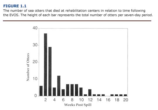
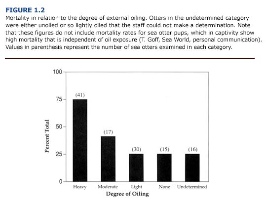
Even with extensive postmortem examination and tissue analysis, it is difficult to differentiate between the pathological effects of hypothermia, stress, and petroleum hydrocarbon toxicity for oiled otters. Mortality can result from a combination of these factors. Consideration must be given to the direct effects of oil contamination as well as to indirect effects associated with the rehabilitation process (i.e. stress of capture, transport, and captivity). The mortality rate for adult sea otters will also depend on degree of oiling and duration of contamination following a spill. During the first three weeks following the EVOS, over 60% of the sea otters arriving at rehabilitation centers died; mortality declined sharply after this initial period (Figure 1.1). Overall, 34% of the sea otters brought to rehabilitation centers died. Heavily oiled otters showed the highest mortality (75 %). By comparison, 41% of the moderately oiled and 25% of the lightly oiled otters died (Figure 1.2). A 25% mortality rate also occurred for sea otters that were unoiled or for which the degree of oiling could not be determined. The similarity in mortality rates for lightly oiled, unoiled, and unclassified animals suggests that factors other than oil exposure contributed to mortality in minimally contaminated animals.
The physical and chemical effects of oil contamination on sea otters will vary according to the chemical composition of oil and the duration of the spill. In an acute event such as the EVOS, the otters’ response to contamination changed as the fresh crude oil weathered during the first two to three weeks (Chapter 4). In a chronic situation, such as long-term exposure in harbors, marine terminals, areas of natural oil seeps, or an oil platform blowout, the effects of contamination may be cumulative.
Physical Effects of Oil
In many ways, the physical effects of oil contamination appear to be more damaging to sea otters than the toxicological effects. Many petroleum products, including fresh crude oil, may contain chemicals that are irritating to the interdigital webbing of the hind flippers and sensitive membranes around the eyes, nose, mouth, and urogenital tissues of the otters. Also, excessive grooming of irritated areas has been found to cause permanent damage including corneal damage, scarring, and alopecia (baldness) (Williams, 1990).
Heavily and moderately oiled sea otters often become hypothermic following contamination of their pelage. (See Chapter 5.) Crude oil rapidly penetrates the fur and destroys its water repellency. This leads to saturation of the pelt and compensatory increases in metabolic rate (Costa and Kooyman, 1982, Williams et al., 1988; Davis et al., 1988). The loss of thermal insulation initiates a suite of physiological, biochemical, and behavioral changes associated with hypothermia. Aside from the mortality directly associated with a decrease in core body temperature, a hypothermic event may also lead to long-term organ damage and dysfunction due to vascular system collapse and congestion. In humans and experimental animals, sudden decreases in core body temperature (acute hypothermia) result in decreased cardiac function, circulatory collapse, and severe congestion in many organs (Paton, 1991). Vascular congestion can lead to hypoxia and ischemic necrosis in organs such as the liver, kidneys, brain, and lungs. The extent of tissue damage will depend on the duration and severity of the hypoxia. An oiled animal may survive the hypothermic event, but suffer impaired organ function as a result of hypoxic damage.
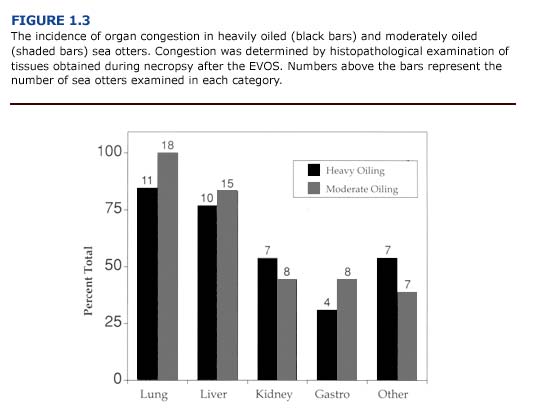
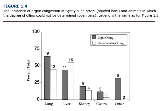
Vascular congestion within specific organs was evident in many of the sea otters that died during the EVOS. The incidence of congestion was site specific and depended on the degree of external oiling. Thus, tissues from heavily and moderately oiled otters (Figure 1.3) were more likely to exhibit congestion than those from lightly or unoiled otters (Figure 1.4). The most prevalent sites of congestion were the lungs and liver. More than 77% of the heavily and moderately oiled sea otters showed hepatic congestion, and nearly 85% showed congestion in the lungs. By comparison, 55% of the lightly oiled and unoiled otters showed hepatic congestion, and 45-64% exhibited lung congestion.
In humans, pancreatitis and gastric hemorrhages are considered characteristic lesions of hypothermia (Paton, 1991).If the patient survives, elevated serum amylase and superficial ulceration (erosion) of the stomach mucosa may develop. Pancreatic injury was not observed in oiled sea otters. However, gastrointestinal hemorrhaging and ulceration occurred frequently and correlated with the degree of external oiling. Similar to findings for hypothermic humans, the gastrointestinal lesions of oiled sea otters were located primarily in the stomach, and occasionally in the duodenum (Williams and Davis, 1990). Gastric erosions may also occur in stressed sea otters, whether the animal is hypothermic or not. In view of this, and because of the focal nature of the lesions, it is likely that the combined effects of hypothermia, stress, shock, captivity, and oil contamination rather than oil ingestion per se were the primary factors leading to gastrointestinal injury in these animals (Lipscomb et al., 1993, 1994).
Chemical Effects of Oil
The toxicological effects of oil contamination are not fully understood for marine mammals. This is due in part to uncertainties about the duration, degree, and route (inhalation, trans dermal, oral) of exposure during an oil spill. When phocid seals were placed in oil-covered water or fed small quantities of crude oil, gastrointestinal irritation, renal tubular necrosis, and liver degeneration were observed (Smith and Geraci, 1975; Engelhardt et al., 1977; Geraci and St. Aubin, 1990). In polar bears, immunosuppression and anemia were also observed (%D8ritsland et al., 1981).
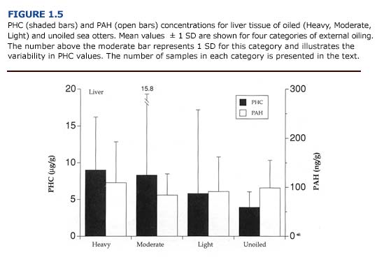
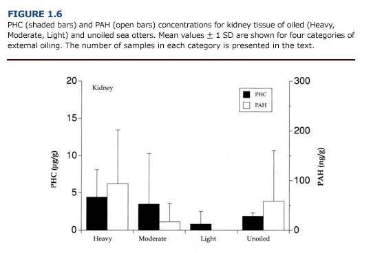
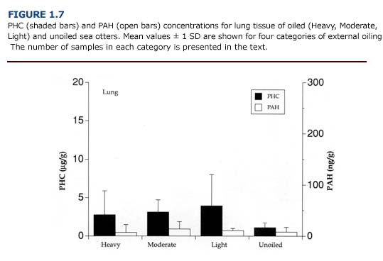
Following the EVOS, the concentration of petroleum hydrocarbons (PAH, PHC) in tissues was highly variable in oiled sea otters (Figures 1.5, 1.6, 1.7). In general, tissue PHC and PAH levels correlated poorly with the degree of external oiling (2 way ANOVA, PHC: F3,53 = 0.36, p= 0.78; PAH: F3,52= 0.35, p = 0.79). Animals from all oiling categories had elevated petroleum hydrocarbons in their tissues. In contrast, PHC and PAH levels varied significantly with tissue type (2 way ANOVA, PHC: F2,53 = 4.69, p%26lt;0.0l; PAH: F2,52= 24.02, p%26lt;0.00l; tissue x oil: F6,56 = 0.94, p = 0.48). With the exception of one unoiled animal, only heavily oiled sea otters showed elevated petroleum hydrocarbon levels in two or more organs.